Pancreaticoduodenectomy for borderline resectable pancreatic head cancer with a modified artery-first approach technique
Min Wang, Hang Zhang, Feng Zhu, Feng Peng, Xin Wang, Ming Shen and Ren-Yi Qin
Wuhan, China
Pancreaticoduodenectomy for borderline resectable pancreatic head cancer with a modified artery-first approach technique
Min Wang, Hang Zhang, Feng Zhu, Feng Peng, Xin Wang, Ming Shen and Ren-Yi Qin
Wuhan, China
BACKGROUND: The treatment of borderline resectable pancreatic head cancer (BRPHC) is still controversial and challenging. The artery-first approaches are described to be the important options for the early determination. Whether these approaches can achieve an increase R0 rate, better bleeding control and increasing long-term survival for BRPHC are still controversial. We compared a previously reported technique, a modified artery-first approach (MAFA), with conventional techniques for the surgical treatment of BRPHC.
METHODS: A total of 117 patients with BRPHC undergone pancreaticoduodenectomy (PD) from January 2013 to June 2015 were included. They were divided into an MAFA group (n=78) and a conventional-technique group (n=39). Background characteristics, operative data and complications were compared between the two groups.
RESULTS: Mean operation time was significantly shorter in the MAFA group than that in the conventional-technique group (313 vs 384 min; P=0.014); mean volume of intraoperative blood loss was significantly lower in the MAFA group than that in the conventional-technique group (534 vs 756 mL; P=0.043); and mean rate of venous resection was significantly higher in the conventional-technique group than that in the MAFA group (61.5% vs 35.9%; P=0.014). Pathologic data, early mortality and morbidity were not different significantly between the two groups.
CONCLUSIONS: MAFA is safe, simple, less time-consuming, less intraoperative blood loss and less venous resection, and therefore, may become a standard surgical approach to PD for BRPHC with the superior mesenteric vein-portal vein involvement but without superior mesenteric artery invasion.
(Hepatobiliary Pancreat Dis Int 2017;16:215-221)
pancreatic head cancer;
pancreaticoduodenectomy;
borderline resectable
Introduction
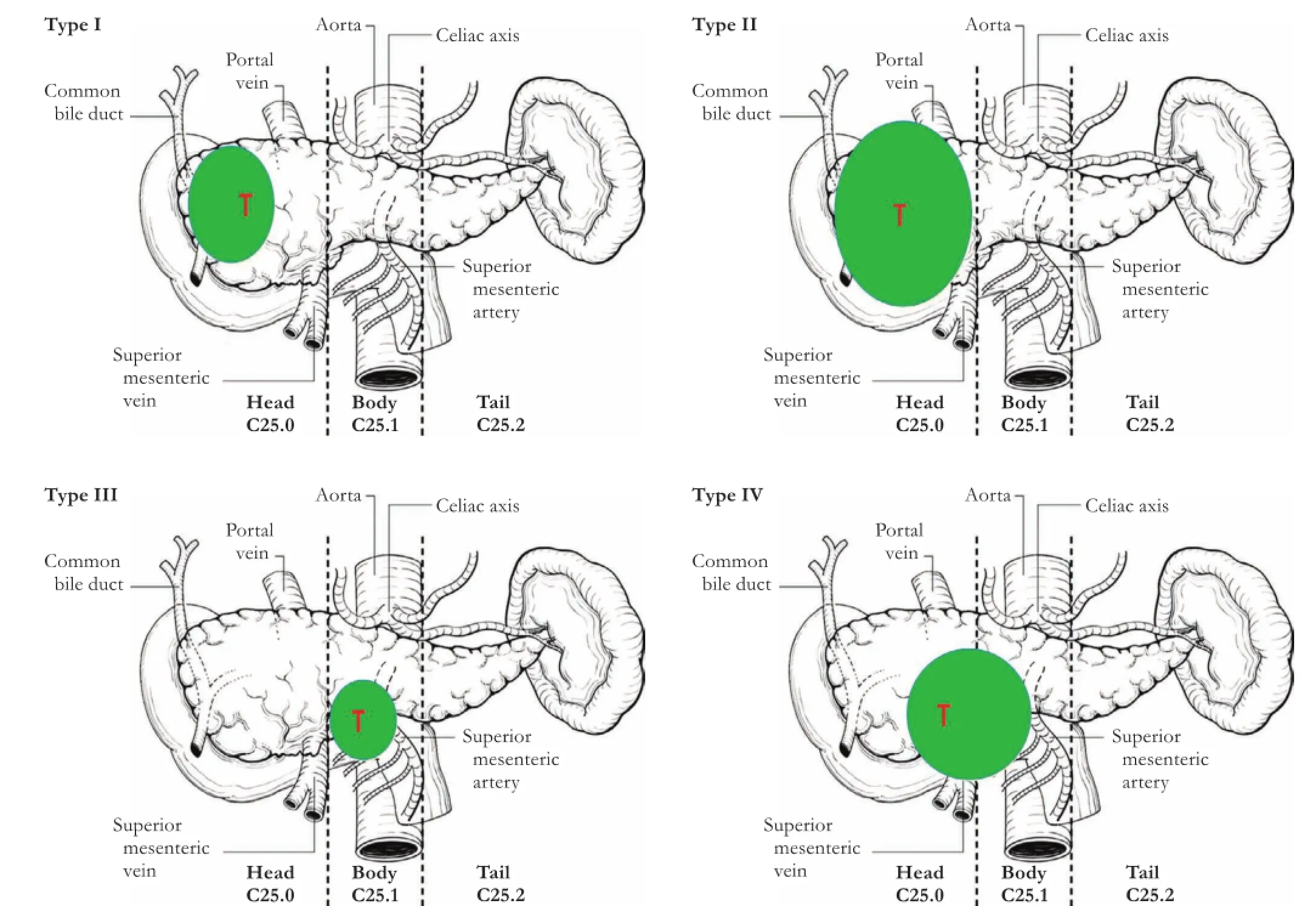
Surgery is the only possible cure treatment for patients with pancreatic head cancer, and achieving a negative-margin (R0) resection is critical to longterm survival.[1-3]The mortality following pancreaticoduodenectomy (PD) has been a remarkable decline with the advances of surgical technique.[2]However, 80% of patients with pancreatic cancer had concomitant distant metastases at the time of diagnosis and only 10% were clearly resectable, and 10% were potentially inoperable because of the advanced stage.[4]This has driven surgeons to improve resection rates. A consensus on the definition and treatment of borderline resectable pancreatic head cancer (BRPHC) was published by the International Study Group of Pancreatic Surgery (ISGPS).[5]During the last decade, vascular resection tecniques used in borderline resectable pancreatic cancer have remarkably improved resection rates.[6,7]The ISGPS supports the imaging-based National Comprehensive Cancer Network (NCCN) criteria for borderline resectability in cases of venous mesentericoportal-axis involvement and arterial involvement.[5]We propose to categorize pancreatic head cancer into four distinct types based on vascular involvement. Type I is pancreatic head cancer without vascular invasion; based on NCCN criteria, it is considered a localized and clearly resectable pancreatic head cancer. Types II, III, and IV pancreatic head cancer are BRPHCas defined by the ISGPS.[5]Type II patients have BRPHC with venous-only mesentericoportal-axis involvement; type III, BRPHC with arterial-only involvement; and type IV, BRPHC with both venous and arterial involvement. In pancreatic head cancer, venous mesentericoportal-axis involvement is most common in BRPHC (Fig. 1).
Recent improvements of surgical technique and perioperative clinical care have reduced the morbidity and mortality after PD. However, PD for pancreatic head cancer, especially borderline resectable, remains a substantial challenge for most surgeons.[8]A safe and effective surgical technique designed especially for BRPHC is warranted to improve resection rates.[6]The criterion for BRPHC is whether the vasculature around the pancreas is involved. The most time-consuming and demanding step during pancreatic resection is dissection of this vasculature, which can result in severe intraoperative and postoperative bleeding.[9]The complexity of this portion of the procedure is demonstrated by the continuing efforts of many researchers to simplify it while maintaining its safety, which has resulted in such techniques as the artery-first approach,[10]no-touch isolation technique,[11]uncinate process-first approach,[12,13]and the hanging maneuver.[14,15]However, there is no agreement as to which of these techniques should be used.[16]
Until now, six artery-first approaches were reported which provide some options to start the surgery. Almost all of the techniques are aimed to determine resectability, not to control the bleeding. In a previous paper, we introduced a modified artery-first approach (MAFA), named“total arterial devascularization-first (TADF)” technique. The basic principle of TADF is first to divide all arteries feeding the tumor in type II BRPHC.[17]In the present study, we retrospectively compared patients characteristics, operative data, and short-term postoperative outcomes of this technique with those of conventional techniques that use vein-first or conventional artery-first approaches.

Fig. 1. Representative pictures of classification of pancreatic head cancer. Type I: pancreatic head cancer without any vascular invasion (SMV/PV, SMA, HA, SA, SV, or CA); Type II: pancreatic head cancer with only SMV/PV invasion, but not SMA invasion; Type III: pancreatic head cancer without SMV/PV invasion or compression, but suppression or invasion of the SMA (<180 degrees) is present; Type IV: pancreatic head cancer with both SMV/PV and SMA involvement or compression (<180 degrees). CA: celiac axis; HA: hepatic artery; PV: portal vein; SA: splenic artery; SMA: superior mesenteric artery; SMV: superior mesenteric vein; SV: splenic vein; T: tumor.
Methods
Patients
A total of 117 patients with BRPHC undergone PD from January 2013 to June 2015 at our institution were included in this study. Patients were divided into two groups: those who underwent PD using the MAFA technique (MAFA group, n=78) from February 2014 to June 2015 and those who underwent PD using conventional techniques (uncinate process-first approach, n=11; veinfirst approach, n=19; or artery-first approach, n=9) from January 2013 to January 2014.
This study only included patients with BRPHC with venous-only mesentericoportal-axis involvement (type II); that is, without distant metastases, with venous involvement of the superior mesenteric vein (SMV) or portal vein (PV) with distortion or narrowing of the vein or occlusion of the vein, and with suitable vessel proximally and distally, allowing for safe resection and replacement. Excluding criteria: 1) patients had severe complications, such as cardiovascular disease, respiratory disease, hepatic and renal dysfunction, stroke, et al; 2) patient age was over 75 years old; and 3) venous occlusion was more than 3 cm and feasible for operation. None patient received neoadjuvant chemotherapy.
Patient characteristics [age, gender, body mass index (BMI), preoperative total bilirubin, type and classification of the infiltrated veins], intraoperative data (operation duration, blood loss, intraoperative RBC transfusion, and type of vein resection); histological diagnosis, and pathological data, including resection margins, T stage, vein and lymph node positive rate, and patient outcomes (in-hospital postoperative mortality and morbidity and postoperative length of stay), were prospectively recorded.
All patients underwent Gem energy spectrum CT with thin-slice scans and vessel reconstruction. A detailed assessment of resectability was carried out by two radiologists using criteria that followed NCCN Guidelines®, Version 1.2013. Resected-margin status was microscopically evaluated by intra- and postoperative pathologic examination. The incidence of postoperative pancreatic fistula, classified into three groups (A, B, and C) according to the International Study Group of Pancreatic Fistula (ISGPF) criteria, was recorded. Delayed gastric emptying, post-pancreatectomy hemorrhage, and biliary fistula were graded according to the ISGPS definitions.[18,19]Operative mortality was defined as any death that occurred within 30 days of the procedure. In this study, all procedures were performed without pyloruspreserving radical PD as previously described.[17]
Surgical technique
The modified artery-first approach
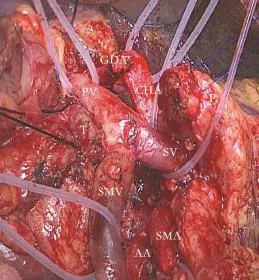
The MAFA has been described in detail in our previous paper (named TADF technique).[17]The basic principle is to isolate all arteries feeding the tumor before treating the involved veins in type II BRPHC. Initially or later in the procedure, a Kocher maneuver is performed to clear the lymph node of the retroperitoneum and expose the anterior surface of the inferior vena cava, and the left side of the aorta, thus enabling dissection of all posterior attachments to the pancreatic head. Then we interrupt arterial blood flow to the region of the pancreatic head in two locations via lymphadenectomy of the hepatic arteries and hepatoduodenal ligament and transection of the pancreatic neck. The right gastric artery and the gastroduodenal artery are ligated and cut after the common hepatic artery (HA) and proper HA dissected and slung. In this approach, we dissect the attachment between the pancreatic mesouncinate and superior mesenteric artery (SMA)/celiac trunk, which completely stops all arterial flow to the pancreatic head. After all arteries of the pancreatic head stop, the specimen is just attached to the right wall of the SMV-PV. All veins (SMV, PV, splenic vein and inferior mesenteric vein) are controlled by three or four vascular-blocking bands. The involved SMV-PV is clamped and resected and then reconstructed[17](Fig. 2).
Conventional technique

Fig. 2. Key steps in the MAFA technique. 1) Exposure of the anterior surface of the abdominal aorta by completely transecting neural and connective tissues between the SMA and pancreatic mesouncinate; 2) Transection of the mesouncinate from the origin of the SMA to the root of the celiac trunk. With these steps, all arterial blood supply to the pancreatic head is interrupted, and three vascular blocking bands for the SMV, PV, and SV were preset to control all venous blood supply before dissection of veins. AA: abdominal aorta; CHA: common hepatic artery; GDA: gastroduodenal artery; P: pancreatic stump; PV: portal vein; SMA: superior mesenteric artery; SMV: superior mesenteric vein; SV: splenic vein; T: tumor.
The conventional techniques used were quite simi-lar to those practiced in most surgical centers and are described elsewhere. Briefly, the Kocher maneuver is performed; the gastrocolic omentum is isolated, the common bile duct is ligated and isolated and the stomach is transected. Following these isolations, the anterior surface of the SMV-PV is identified. The right gastric artery and the gastroduodenal artery were separated. After complete detachment of the PV from the posterior surface of the pancreatic neck, the pancreas is transected. The involved SMV-PV is clamped and resected and then reconstructed (with or without graft interposition, depending on the local situation). In this group, we also use some artery-first approaches, the no-touch isolation technique, the uncinate process-first approach, and the hanging maneuver. However, these techniques do not block the arterial flow to the pancreatic head before treating the involved veins.[10,12,14,20]
Statistical analysis
SPSS Statistics for Windows, Version 17.0 (SPSS Inc., Chicago, IL, USA) was used for data analysis. For continuous variables, descriptive statistics were calculated and reported as the mean±standard deviation (SD). Categorical variables were described using frequency distributions. An independent-sample t test was used to detect differences between the means of continuous variables, and the Chi-square test was used in cases with low expected frequencies. P values <0.05 were considered statistically significant.
Results
Patient characteristics
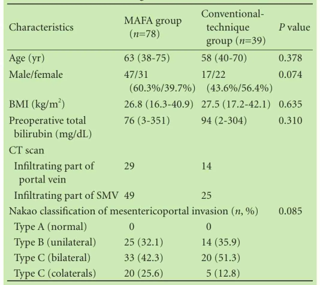
From January 2013 to June 2015, of the 117 consecutive patients with BRPHC underwent radical PD, 78 underwent MAFA, and the other 39 underwent the conventional techniques. The preoperative data, including age, gender, BMI, and preoperative serum bilirubin concentration in the two groups were comparable (Table 1). There was no significant difference between groups in the degree of venous invasion, as assessed using the classification system by Nakao et al.[21]
Operative data
Mean operative duration was significantly shorter in the MAFA group than that in the conventionaltechnique group [313 (220-439) vs 384 (245-502) min, P=0.014]. Mean volume of intraoperative blood loss was significantly lower in the MAFA group than that in the conventional-technique group [534 (300-1400) vs 756 (320-3300) mL, P=0.043]. No serious intraoperative incident occurred in either group (Table 2).
The rate of venous resection was significantly lower in the MAFA group than that in the conventional-technique group (35.9% vs 61.5%, P=0.014). In the MAFA group, 9 patients underwent PV resection and 19 underwent SMV resection; in the conventional-technique group, 9 underwent PV resection and 15 underwent SMV resection. In the MAFA group, 21 underwent vessel lateral wall resection and 7 underwent segmental vessel resection and end-to-end anastomosis; in the conventional-technique group, 12 patients underwent vessel lateral wall resection and 12 underwent segmental vessel resection and end-to-end anastomosis. Graft interposition was used in only one patient in the conventionaltechnique, for venous reconstruction.
Pathologic data
There were no significant differences between groups in grade of differentiation, T stage, and tumor vascular invasion. There were no significant differences in surgical margin status and number of positive lymph nodes dissected between the two groups. The number of patients with microscopic residual tumor did not differ between the MAFA (9, 11.54%) and conventional technique groups (4, 10.25%; P=0.53) (Table 2).
Early mortality and morbidity
Surgical outcomes and postoperative complications are presented in Table 2. There were no postoperative deaths in either group. There was no significant differ-ence in complication rate between groups (P=0.603). Postoperative major complications were encountered in 32 patients in the MAFA group (41.0%) and 18 patients in the conventional-technique group (46.2%). All postoperative pancreatic fistula were conservatively managed by maintaining the closed suction drains and using octreotide without additional intervention. Other complications were managed in all patients nonoperatively, by maintenance or reinsertion of a nasogastric tube, treating with antibiotics and parenteral nutrition.

Table 1. Background characteristics

Table 2. Surgical outcomes and postoperative complications
There was no significant difference in length of postoperative hospitalization between groups (P=0.360), and no patient in either group died of tumor recurrence around SMV/SMA within 3 months after the operation.
Discussion
In the present study, we compared the results of PD with MAFA to those of PD using conventional approaches for BRPHC. Preliminary results showed that MAFA for type II BRPHC significantly reduced intraoperative bleeding and operative duration.
To guide operative decisions, we suggest classifying pancreatic head cancer by the relationship of the tumor to key vasculature around the pancreatic head. Several classification systems, described different aspect of the tumor, have been used to describe the radiologic and prognosis appearance of pancreatic cancer, such as the Union Internationale Contre le Cancer (UICC) and Japan Pancreas Society (JPS) classifications.[22]However, despite the worldwide application of these systems, little information on their use in operative decision-making is available. We believe there is a need for a classification system for pancreatic cancer that will assist operative decision-making and that different types of BRPHC should use different techniques.
The golden standard surgical procedure for pancreatic head cancer is still controversial. The key problem associated with these techniques is that they involve one method for addressing all types of pancreatic cancer. The traditional techniques included the no-touch technique, uncinate process-first approach, vein-first approach, and artery-first approach. However these methods used for BRPHC always cause serious intraoperative bleeding. Hirota et al[11]introduced a no-touch technique that involves hanging and clamping. This technique has many potential advantages, including no squeezing of the pancreas by the surgeon and achievement of R0 resection. However, there is a high risk associated with first treating the SMV-PV in type I BRPHC patients. The arteryfirst approach is also an important and useful technique for this type of pancreatic head cancer. Although severalartery-first approaches are considered especially helpful in patient with locally advanced tumor, no approach completely halts arterial blood flow to the pancreatic head before the involved veins are treated.[10]In contrast to the artery-first approaches for early determination of the SMA, here we have not only explored the SMA, but also emphasized completely blocking arterial blood flow to the pancreatic head before treating the involved veins. The conventional techniques emphasize the block of the blood supplies from HA and splenic artery by dividing the gastroduodenal artery and pancreas. However, the blood supplies from SMA and arteries of pancreatic mesouncinate always are spared. The critical steps of the MAFA technique in this study are to completely transect neural and connective tissues between SMA and pancreatic mesounsinate, and transecting the mesounsinate from the origin of SMA to the root of the celiac trunk.[17]After innovatively handling the arterial vessels of the pancreatic head first, there is no arterial blood supply, and bleeding of the specimen is thus reduced. With vascular-blocking bands, the veins can be controlled, and so the venous bleeding, when treating the involved veins. The MAFA technique can reduce intraoperative blood loss by allowing precise control of the blood supplies to the pancreatic head.
It is still difficult to distinguish the difference between non-neoplastic inflammatory infiltration and true malignant infiltration leading to non-validated, individualized treatment regimens.[23]Therefore, vascular resection and reconstruction at the time of PD is still controversial.[24]One of the largest systematic reviews on this topic, published in 2006 and including 1646 patients with PV and/or SMV resection from 52 studies, indicated that PV resection did not improve survival rates over non-resective treatments.[24]However, another study reported that morbidity, mortality, and disease-free and overall survival were the same as those of patients who underwent standard resection and better than those of patients who did not undergo operative treatment because of venous involvement.[25]Based on these data, the limiting factor for resectability where there is venous involvement is therefore the possibility of venous reconstruction. However, it may not be prudent to remove so many veins without evidence of true tumor involvement rather than inflammatory infiltration.[26,27]In MAFA technique, we first blocked all arterial blood flow and used vascular-blocking bands to control all venous flow from the pancreatic head. By precisely controlling blood flow, the involved veins could be safely dissected. However, by the conventional techniques, the arteries are not completely isolated before the venous resection. If we want to divide the attachment between the pancreatic mesonucinate and SMA, we should remove the involved veins to expose the pancreatic mesonucinate. During this process, it is very dangerous to dissect the involved veins, so we do more veins resection, but not dissection. In the present study, most involved veins could be dissected without vein resection by the MAFA technique; we then sent the dissected specimen for intraoperative frozen section to assist with decision-making regarding vein resection. In this way we were able to achieve the same R0 rate with less vein resection. By employing the MAFA technique, needless vein resection is significantly reduced.
Because this work is preliminary and retrospective in one group, two limitations may cause the bias for our conclusions. One is that the time periods did not overlap. Our outcome cannot exclude the effect of the improved technique of the surgeon over time on patients’ outcomes. Second we have not presented the oncologic/survival data for the short follow-up time. The deficiency of the oncologic/survival may confuse the effect of the low rate of the vein resection by the new technique. So the technique must be evaluated in larger RCTs to evaluate overall and oncologic/survival data in the future.
In conclusions, we developed a classification system for pancreatic head cancer and employed MAFA surgical technique for precise control of blood flow in the pancreatic head region. The MAFA technique can improve surgical safety, reduce the incidence of postoperative bleeding, and achieve the no-touch principle via control of blood flow. Our data suggest that MAFA is a safe and effective surgical method for resection of BRPHC. MAFA has the potential to become a standard surgical approach for intricate radical PD, especially in patients with SMA/ PV invasion.
Contributors: WM drafted the article, analyzed and interpreted the data. ZH collected, analyzed, and interpreted the data. PF collected the data. ZF, WX and SM designed the study and collected the data. QRY designed the study and critically revised the article. QRY is the guarantor.
Funding: This study was supported by grants from The National Natural Science Foundation of China (81071775, 81272659, 81101621, 81172064, 81001068 and 81272425), Key Projects of Science Foundation of Hubei Province (2011CDA030), and Research Fund of Young Scholars for the Doctoral Program of Higher Education of China (20110142120014).
Ethical approval: This study was approved by the Ethics Committee of the Tongji Hospital, Tongji Medical College, Huazhong University of Science and Technology.
Competing interest: No benefits in any form have been received or will be received from a commercial party related directly or indirectly to the subject of this article.
1 Hidalgo M. Pancreatic cancer. N Engl J Med 2010;362:1605-1617.
2 La Torre M, Nigri G, Ferrari L, Cosenza G, Ravaioli M, Ramacciato G. Hospital volume, margin status, and long-term survival after pancreaticoduodenectomy for pancreatic adenocarcinoma. Am Surg 2012;78:225-229.
3 Cameron JL, Riall TS, Coleman J, Belcher KA. One thousand consecutive pancreaticoduodenectomies. Ann Surg 2006;244: 10-15.
4 Vincent A, Herman J, Schulick R, Hruban RH, Goggins M. Pancreatic cancer. Lancet 2011;378:607-620.
5 Bockhorn M, Uzunoglu FG, Adham M, Imrie C, Milicevic M, Sandberg AA, et al. Borderline resectable pancreatic cancer: a consensus statement by the International Study Group of Pancreatic Surgery (ISGPS). Surgery 2014;155:977-988.
6 Katz MH, Crane CH, Varadhachary G. Management of borderline resectable pancreatic cancer. Semin Radiat Oncol 2014;24: 105-112.
7 Ravikumar R, Sabin C, Abu Hilal M, Bramhall S, White S, Wigmore S, et al. Portal vein resection in borderline resectable pancreatic cancer: a United Kingdom multicenter study. J Am Coll Surg 2014;218:401-411.
8 Yamada S, Fujii T, Sugimoto H, Nomoto S, Takeda S, Kodera Y, et al. Aggressive surgery for borderline resectable pancreatic cancer: evaluation of National Comprehensive Cancer Network guidelines. Pancreas 2013;42:1004-1010.
9 Sharma J, Ng J, Goodman MD, Saif MW. New developments in the management of borderline resectable pancreatic cancer. JOP 2013;14:123-125.
10 Weitz J, Rahbari N, Koch M, Büchler MW. The “artery first”approach for resection of pancreatic head cancer. J Am Coll Surg 2010;210:e1-4.
11 Hirota M, Kanemitsu K, Takamori H, Chikamoto A, Tanaka H, Sugita H, et al. Pancreatoduodenectomy using a no-touch isolation technique. Am J Surg 2010;199:e65-68.
12 Hackert T, Werner J, Weitz J, Schmidt J, Büchler MW. Uncinate process first--a novel approach for pancreatic head resection. Langenbecks Arch Surg 2010;395:1161-1164.
13 Shrikhande SV, Barreto SG, Bodhankar YD, Suradkar K, Shetty G, Hawaldar R, et al. Superior mesenteric artery first combined with uncinate process approach versus uncinate process first approach in pancreatoduodenectomy: a comparative study evaluating perioperative outcomes. Langenbecks Arch Surg 2011;396:1205-1212.
14 Addeo P, Marzano E, Rosso E, Pessaux P. Hanging maneuver during pancreaticoduodenectomy: a technique to improve R0 resection. Surg Endosc 2011;25:1697-1698.
15 Marzano E, Piardi T, Pessaux P. The “hanging maneuver” technique during pancreaticoduodenectomy: the result of a technical evolution to approach the superior mesenteric artery. JOP 2011;12:429-430.
16 Katz MH, Marsh R, Herman JM, Shi Q, Collison E, Venook AP, et al. Borderline resectable pancreatic cancer: need for standardization and methods for optimal clinical trial design. Ann Surg Oncol 2013;20:2787-2795.
17 Peng F, Wang M, Zhu F, Tian R, Shi CJ, Xu M, et al. “Total arterial devascularization first” technique for resection of pancreatic head cancer during pancreaticoduodenectomy. J Huazhong Univ Sci Technolog Med Sci 2013;33:687-691.
18 Bassi C, Dervenis C, Butturini G, Fingerhut A, Yeo C, Izbicki J, et al. Postoperative pancreatic fistula: an international study group (ISGPF) definition. Surgery 2005;138:8-13.
19 Wente MN, Bassi C, Dervenis C, Fingerhut A, Gouma DJ, Izbicki JR, et al. Delayed gastric emptying (DGE) after pancreatic surgery: a suggested definition by the International Study Group of Pancreatic Surgery (ISGPS). Surgery 2007;142:761-78.
20 Shukla PJ, Barreto G, Pandey D, Kanitkar G, Nadkarni MS, Neve R, et al. Modification in the technique of pancreaticoduodenectomy: supracolic division of jejunum to facilitate uncinate process dissection. Hepatogastroenterology 2007;54:1728-1730.
21 Nakao A, Kanzaki A, Fujii T, Kodera Y, Yamada S, Sugimoto H, et al. Correlation between radiographic classification and pathological grade of portal vein wall invasion in pancreatic head cancer. Ann Surg 2012;255:103-108.
22 Kawarada Y. JPS 5th ed. Classification of pancreatic cancer and JPS classification versus UICC classification. Nihon Rinsho 2006;64:81-86.
23 Tempero M, Arnoletti JP, Ben-Josef E, Bhargava P, Casper ES, Kim P, et al. Pancreatic adenocarcinoma. Clinical practice guidelines in oncology. J Natl Compr Canc Netw 2007;5:998-1033.
24 Siriwardana HP, Siriwardena AK. Systematic review of outcome of synchronous portal-superior mesenteric vein resection during pancreatectomy for cancer. Br J Surg 2006;93:662-673.
25 Adham M, Mirza DF, Chapuis F, Mayer AD, Bramhall SR, Coldham C, et al. Results of vascular resections during pancreatectomy from two European centres: an analysis of survival and disease-free survival explicative factors. HPB (Oxford) 2006;8:465-473.
26 Tseng JF, Raut CP, Lee JE, Pisters PW, Vauthey JN, Abdalla EK, et al. Pancreaticoduodenectomy with vascular resection: margin status and survival duration. J Gastrointest Surg 2004;8:935-950. 27 Nakagohri T, Kinoshita T, Konishi M, Inoue K, Takahashi S. Survival benefits of portal vein resection for pancreatic cancer. Am J Surg 2003;186:149-153.
Received July 22, 2015
Accepted after revision September 30, 2016
Author Affiliations: Department of Pancreatic-Biliary Surgery, Affiliated Tongji Hospital, Tongji Medical College, Huazhong University of Science and Technology, Wuhan 430030, China (Wang M, Zhang H, Zhu F, Peng F, Wang X, Shen M and Qin RY)
Ren-Yi Qin, MD, Department of Biliary-Pancreatic Surgery, Affiliated Tongji Hospital, Tongji Medical College, Huazhong University of Science and Technology, 1095 Jiefang Ave., Wuhan 430030, China (Tel/Fax: +86-27-8366-5294; Email: ryqin@tjh.tjmu.edu.cn)
© 2017, Hepatobiliary Pancreat Dis Int. All rights reserved.
10.1016/S1499-3872(16)60171-6
Published online February 24, 2017.
 Hepatobiliary & Pancreatic Diseases International2017年2期
- Hepatobiliary & Pancreatic Diseases International的其它文章
- Long-term outcome of patients with chronic pancreatitis treated with micronutrient antioxidant therapy
- High-grade pancreatic intraepithelial lesions: prevalence and implications in pancreatic neoplasia
- HIDA scan for functional gallbladder disorder: ensure that you know how the scan was done
- Novel HBV mutations and their value in predicting efficacy of conventional interferon
- Serum soluble ST2 is a promising prognostic biomarker in HBV-related acute-on-chronic liver failure
- The association of non-alcoholic fatty liver disease and metabolic syndrome in a Chinese population
