The relationship between SPARC expression in primary tumor and metastatic lymph node of resected pancreatic cancer patients and patients' survival
Shanghai, China
The relationship between SPARC expression in primary tumor and metastatic lymph node of resected pancreatic cancer patients and patients' survival
Xin-Zhe Yu, Zhong-Yi Guo, Yang Di, Feng Yang, Qi Ouyang, De-Liang Fu and Chen Jin
Shanghai, China
BACKGROUND: Previous researches in pancreatic cancer demonstrated a negative correlation between secreted protein acidic and rich in cysteine (SPARC) expression in primary tumor and survival, but not for SPARC expression in lymph node. In the present study, we aimed to evaluate the SPARC expression in various types of tissues and its impact on patients' prognosis.
METHODS: The expression of SPARC was examined by immunohistochemistry in resected pancreatic cancer specimens. Kaplan-Meier analyses and Cox proportional hazards regression were applied to assess the mortality risk.
RESULTS: A total of 222 tissue samples from 73 patients were collected to evaluate the SPARC expression, which included 73 paired primary tumor and adjacent normal tissues, 38 paired metastatic and normal lymph nodes. The proportion of positive SPARC expression in metastatic lymph node was high (32/38), whereas in normal lymph node it was negative (0/38). Positive SPARC expression in primary tumor cells was associated with a signifcantly decreased overall survival (P=0.007) and disease-free survival (P=0.003), whereas in other types oftissues it did not show a predictive role for prognosis. Univariate and multivariate analyses both confrmed this signifcance.
CONCLUSION: SPARC can serve a dual function role as both predictor for prognosis and potentially biomarker for lymph node metastasis in resected pancreatic cancer patients.
(Hepatobiliary Pancreat Dis Int 2017;16:104-109)
pancreatic cancer;
SPARC expression;
immunohistochemistry;
prognostic marker;
lymph node metastasis
Introduction
Pancreatic cancer is the fourth most common cause of cancer death in the Western world.[1]Despite the impressive progress in basic and clinical research, treating pancreatic cancer is still a therapeutic challenge with dismal 5-year survival rates less than 5%.[2,3]In fact, the diffcult surgical approach, resistance to conventional therapies, and the subsequent low survival rate can all contribute to the high-frequency occurrence of lymph node metastasis.[4-6]Thus, fnding an effective biomarker for the lymph node metastasis is critically important.[7]
Secreted protein acidic and rich in cysteine (SPARC), also known as the osteonectin and basement membrane-40 (BM-40) protein, is a member of the matricellular family of proteins.[8,9]SPARC was frst identifed in bone and later in platelets as well as tumor tissues,[2]which plays a pivotal role in cell proliferation,[10]angiogenesis[11]and cell migration.[12]SPARC has been reported to be either up-regulated or down-regulated depending on cancer types,[13]suggesting its complex role in the tumorigenesis. In addition, a hypothesis is proposed that SPARC may act as an albumin “sticker”, which facilitates the deliveryof nab-paclitaxel towards pancreatic cancer cells.[14-16]Recently, SPARC overexpression has been described in colorectal cancer,[17]gastric cancer[18]and pancreatic cancer,[19]and has been associated with poor prognosis.[20-22]Normally, increased SPARC expression has often been found in the stromal area rather than tumor cells, especially in pancreatic cancer.[23]However, no data are available in regards to the SPARC expression in metastatic lymph node and its correlation with survival beneft.
To the best of our knowledge, this study frstly reported data on SPARC expression in lymph node (normal and metastatic) as well as primary tumor of pancreatic cancer patients, and its correlation with the survival beneft. We aimed to explore the dual function role of SPARC as both prognostic predictor and biomarker for the lymph node metastasis in pancreatic cancer patients.
Methods
Clinicopathological data and tissue selection
We retrospectively reviewed 73 patients who underwent radical pancreatic cancer surgery from January 2007 to December 2013 in our hospital. All patients were confrmed to have pancreatic ductal adenocarcinoma by pathologists. Among the 73 patients, 50 were pancreatic head carcinoma, and 23 were pancreatic body or tail carcinoma. A total of 222 tissue samples from these patients were eligible to evaluate the SPARC expression, which included 73 paired primary tumor and matched adjacent normal tissues, 38 paired metastatic and matched normal lymph nodes. All of the clinical and pathologic information was acquired from a regular updated clinical database. The methods for follow-up included, but were not limited to, centers for disease control, outpatient, telephone, and mail. The study was approved by the review board committee of Fudan University.
SPARC immunohistochemistry
Formalin-fxed, paraffn-embedded blocks of tissues from each patient were analyzed with immunohistochemistry, which was performed according to the standard protocol of previous report.[24]Briefy, resected tissues were sliced into 4-5 μm thickness, de-paraffnized, and bathed in citrate buffer at 95 ℃ for 40 minutes for antigen retrieval. The slices were then incubated at 4 ℃for 13 hours with a monoclonal SPARC antibody (Abcam plc, Cambridge, UK), and stained with an avidin-biotin system (Shanghai High-tech Inc., Shanghai, China). All of the nuclei were counterstained with hematoxylin.
SPARC staining analyses were evaluated by two observers (Di Y and Yang F) blinded to the clinical data and treatment arm. Any discrepancy was resolved by a third observer (Ouyang Q). The slices were evaluated for the distribution of staining in the tissues (cytoplasm or stroma). Five microscopic felds from each sample were randomly chosen. The positive cell number and total cell number in every feld were counted. The percentage of positive cell=positive cells/total cells. SPARC immunolabeling was categorized as negative when the intensity was absent to weak (+) and if the extent was less than 10%; immunolabeling was positive if the intensity was moderate (++) to strong (+++), and the extent was ≥10%. SPARC was categorized as positive or negative for the primary tumor site, adjacent normal tissue and lymph node: Tumor-/-Stroma, Tumor+/-Stroma, Tumor-/+Stroma, Tumor+/+Stroma.
Statistical analysis
Overall survival (OS) was defned as the time period from surgery to death by any cause. Disease-free survival (DFS) was defned as the time from surgery to the development of either local recurrence or distant metastasis. The Pearson's Chi-square test or Fisher's exact probability test was used to compare clinicopathological characteristics of patients with positive and negative SPARC expression. Kaplan-Meier analysis and Cox proportional hazards regression modeling were used to assess the mortality risk associated with the presence or absence of tumor SPARC and stromal SPARC status. All of the statistical analyses were performed with SPSS software (version 22.0; IBM Inc., NY, USA). APvalue of <0.05 was considered statistically signifcant.
Results
Patient characteristics
A total of 73 pancreatic cancer patients were analyzed in the pool, with a median follow-up time of 30 months (the cut-off date for follow-up was December, 30th 2014). We collected 73 primary tumor site tissues, 73 adjacent normal tissues, 38 metastatic lymph nodes and 38 matched normal lymph nodes.
SPARC expression in tissues
The SPARC expression was observed both in the cytoplasm of tumor cells and cytoplasm of fbroblasts in the stromal area (Fig. 1). From the results of the immunohistochemistry we found that the SPARC expression was strongly overexpressed in cancer cells and stromal fbroblasts immediately adjacent to cancer cells, suggesting its role in the tumor-stroma interaction. However, SPARC expression was weak or absent in normal pancre-atic ductal cells. Among the 73 tissues from the primary tumor site: twenty-one samples (28.8%) were classifed as Tumor-/+Stroma, and the other 52 samples (71.2%) were classifed as Tumor+/+Stroma; among the 73 tissues from the adjacent normal tissues: ffteen samples (20.5%) were classifed as Stroma+, the other 58 samples (79.5%) were classifed as Stroma-; whereas in 38 tissues from the metastatic lymph node: eight samples (21%) were classifed as Tumor-/+Stroma, six samples (15.8%) were classifed as Tumor-/-Stroma, the other 24 samples (63.2%) were classifed as Tumor+/+Stroma; however, in all tissues from the normal lymph node, there were none SPARC expression. Refer to previous reports,[25-27]we examined SPARC expression in the primary tumor site and its association with major clinicopathological parameters. As shown in Table 1, baseline data and risk factors such as age, gender, tumor size, nodal status, resection margin were comparable between the Tumor-/+Stroma and Tumor+/+Stroma groups.
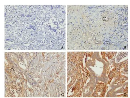
Fig. 1. Patterns of immunohistochemical expression of SPARC in resected pancreatic ductal adenocarcinoma specimens (original magnification ×100). A: Negative staining (-); B: Weakly staining (+); C: Moderately staining (++); D: Strongly staining (+++). ○: positive expression of SPARC.
SPARC expression and patients prognosis
We performed survival analyses in dependence of SPARC expression within all tissue groups. Fig. 2 indicated that negative SPARC expression in tumor cell was associated with a signifcantly increased OS (HR=0.44; 95% CI: 0.24-0.82;P=0.007) and DFS (HR=0.38; 95%CI: 0.20-0.71;P=0.003). However, there seemed no connection between SPARC expression and survival benefts in the metastatic lymph node group (Fig. 3). It was also the same situation in adjacent normal tissue group.
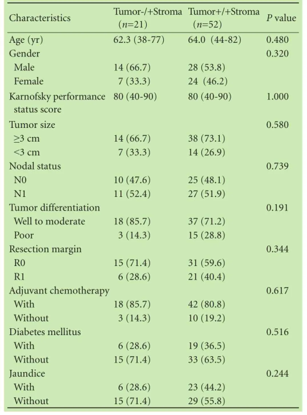
Table 1. Clinicopathological characteristics in relation to SPARC expression in the primary tumor (n, %)

Fig. 2. Kaplan-Meier overall survival curves (A) and disease-free survival curves (B) for the SPARC expression negative vs positive in the primary tumor site.
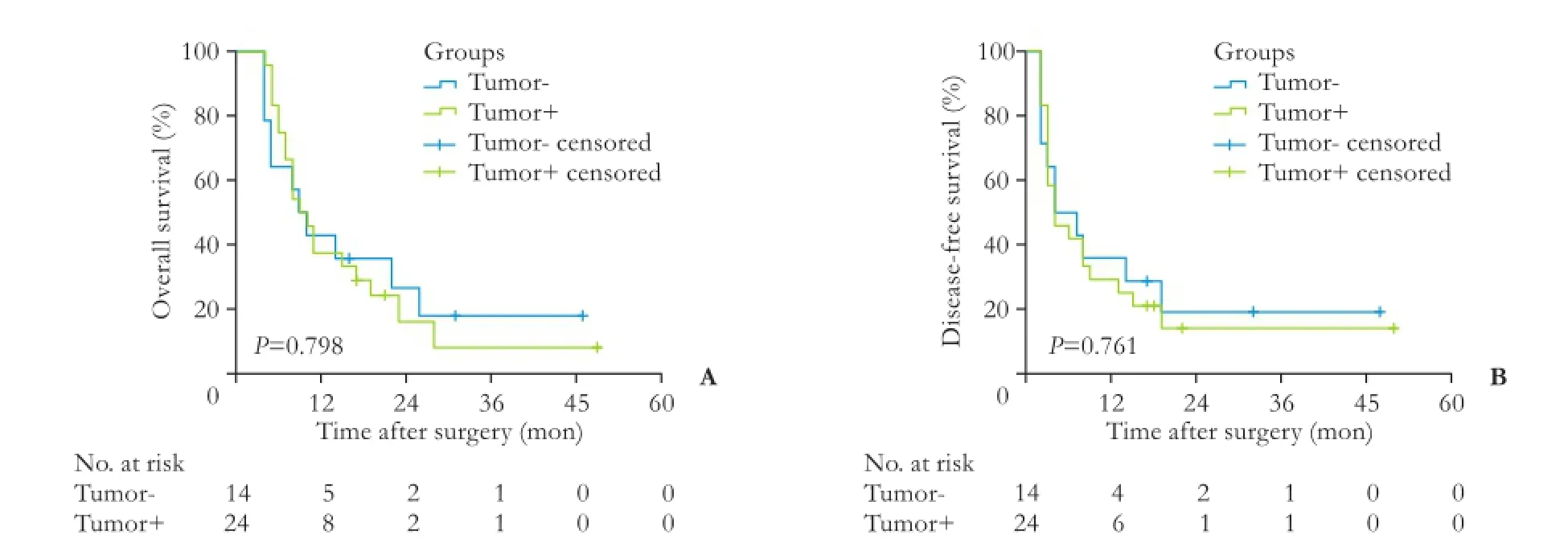
Fig. 3. Kaplan-Meier overall survival curves (A) and disease-free survival curves (B) for the SPARC expression negative vs positive in the metastatic lymph nodes.
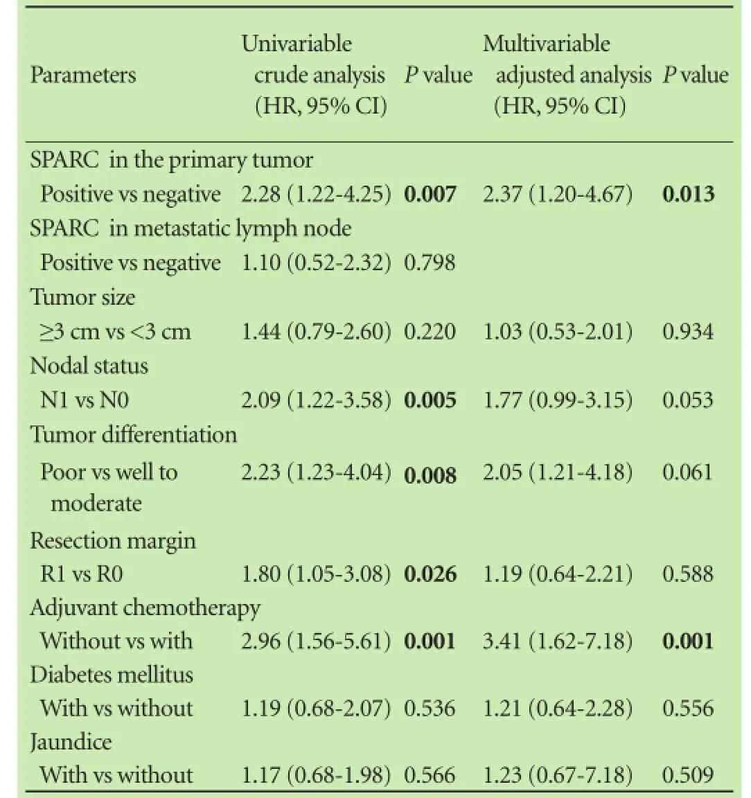
Table 2. Univariate and multivariate analyses of overall survival after surgery
Univariate and multivariate analyses of OS
Univariate analysis revealed that the following parameters were associated with increased mortality: positive SPARC expression in the primary tumor (HR=2.28; 95% CI: 1.22-4.25;P=0.007); metastatic lymph node (HR=2.09; 95% CI: 1.22-3.58;P=0.005); advanced tumor grade (HR=2.23; 95% CI: 1.23-4.04;P=0.008); positive resection margin (HR=1.80; 95% CI: 1.05-3.08;P=0.026); without adjuvant chemotherapy (HR=2.96; 95% CI: 1.56-5.61;P=0.001). In a further step of multivariate analysis, only SPARC expression in the primary tumor (HR=2.37; 95% CI: 1.20-4.67;P=0.013) and without adjuvant chemotherapy (HR=3.41; 95% CI: 1.62-7.18;P=0.001) retained their negative impact on survival (Table 2).
Discussion
SPARC positive or overexpression has often been related to bad prognosis in various malignant tumors,[28-31]several studiesin vivoandin vitroalso confrmed this.[32,33]However, the exact mechanism of action remains unclear.[34,35]
This study shows that positive SPARC expression in the primary tumor cell is associated with a signifcantly decreased OS and DFS, which is generally in accordance with Infante et al's report.[25]In their study, pancreatic cancer patients with positive SPARC expression in primary tumor had a signifcant worse prognosis than those with negative SPARC expression (P<0.001). Notably, this difference was restricted to SPARC expression in the stromal area, they did not fnd a correlation between tumor cell SPARC expression and survival (P=0.13). In another report by Sinn et al,[36]both positive stromal and cytoplasmic SPARC expression was associated with worse DFS and OS. In our study, however, SPARC staining within stromal area was observed in all samples from the primary tumor site; patients with Tumor-/+Stroma had a signifcant longer median survival compared with those with Tumor+/+Stroma. For other type of tissues, including adjacent normal tissues, metastatic lymph nodes, and normal lymph nodes, we did not fnd a correlation between SPARC expression and survival.
Univariate analysis indicated that positive SPARCexpression in the primary tumor, metastatic lymph node, advanced tumor grade, positive resection margin, without adjuvant chemotherapy were all risk factors of overall survival. Multivariate regression analysis showed that positive SPARC expression in the primary tumor was still an independent prognostic factor (HR=2.37; 95% CI: 1.20-4.67;P=0.013) after adjustment for metastatic lymph node, advanced tumor grade, without adjuvant chemotherapy and positive resection margin.
A highlight of this study is that SPARC can potentially serve as a biomarker for the lymph node metastasis. SPARC expression in the metastatic lymph node is higher than that in the matched normal lymph node. Comparing with CA19-9, a generally accepted biomarker for early diagnosis and effcacy evaluation, with a sensitivity of 78.6% and specifcity of 87.3%,[37]SPARC in our study yield a sensitivity of 84.2% (32/38) and specifcity of 100% (38/38). Nevertheless, this result should be treated with caution due to our limited sample size and a retrospective feature. Further validation is needed in a larger cohort study.
For resectable pancreatic cancer patients, chemotherapy always plays a pivotal role. Whether chemotherapy will have an impact on the relationship between SPARC and survival remains controversial. Sinn and coworkers[36]found that the negative impact of SPARC on survival was only restricted to patients who were treated with gemcitabine. Infante et al[25]demonstrated that chemotherapy did not affect the correlation between SPARC and survival.
Our study has several limitations. First one is a retrospective feature with a relatively small size. However, all of the patients and samples were recruited on a consecutive time basis to maximally avoid selection bias. Second, only 38 metastatic lymph nodes were acquired for analysis which may cause type II error. Third, we only used immunohistochemical staining which is not sensitive for protein detection, other methods such as Western blotting and ELISA may improve the sensitivity. Another limitation is that, according to Von Hoff et al,[14]in patients with pancreatic cancer who treated with nab-paclitaxel, SPARC improves chemotherapy response. None of our patient received nab-paclitaxel treatment because nab-paclitaxel is recommended for advanced pancreatic cancer rather than resectable cancer; nab-paclitaxel is expensive and there is no insurance coverage in China now.
In summary, positive SPARC expression predicts poor prognosis, additionally, SPARC has the potential to serve as a biomarker for lymph node metastasis in resected pancreatic cancer patients. Future randomized controlled trials are needed to validate our results.
Contributors:YXZ and DY proposed the study. YXZ, GZY and DY carried out the research. YXZ drafted the manuscript. DY, YF and OQ collected and analyzed the data. FDL and JC kindly offered valuable suggestions about the experiment design. YXZ and GZY contributed equally to this article. JC is the guarantor.
Funding:This study was supported by grants from the National Natural Science Foundation of China (81201896 and 81071884) and the Research Fund for the Doctoral Program of Higher Education of China (20130071110052).
Ethical approval:All specimens were collected in accordance with informed consents of patients, and all procedures complied with the protocol approved by the review board committee of Fudan University.
Competing interest:No benefts in any form have been received or will be received from a commercial party related directly or indirectly to the subject of this article.
1 Siegel R, Ma J, Zou Z, Jemal A. Cancer statistics, 2014. CA Cancer J Clin 2014;64:9-29.
2 Neuzillet C, Tijeras-Raballand A, Cros J, Faivre S, Hammel P, Raymond E. Stromal expression of SPARC in pancreatic adenocarcinoma. Cancer Metastasis Rev 2013;32:585-602.
3 Hidalgo M. Pancreatic cancer. N Engl J Med 2010;362:1605-1617.
4 Yang F, Jin C, Yang D, Jiang Y, Li J, Di Y, et al. Magnetic functionalised carbon nanotubes as drug vehicles for cancer lymph node metastasis treatment. Eur J Cancer 2011;47:1873-1882.
5 Yang F, Jin C, Subedi S, Lee CL, Wang Q, Jiang Y, et al. Emerging inorganic nanomaterials for pancreatic cancer diagnosis and treatment. Cancer Treat Rev 2012;38:566-579.
6 Wagner M, Dikopoulos N, Kulli C, Friess H, Büchler MW. Standard surgical treatment in pancreatic cancer. Ann Oncol 1999;10:247-251.
7 Kim KY, Lee GY, Cha IH. Biomarker detection for the diagnosis of lymph node metastasis from oral squamous cell carcinoma. Oral Oncol 2012;48:311-319.
8 Porter PL, Sage EH, Lane TF, Funk SE, Gown AM. Distribution of SPARC in normal and neoplastic human tissue. J Histochem Cytochem 1995;43:791-800.
9 Motamed K. SPARC (osteonectin/BM-40). Int J Biochem Cell Biol 1999;31:1363-1366.
10 Ryall CL, Viloria K, Lhaf F, Walker AJ, King A, Jones P, et al. Novel role for matricellular proteins in the regulation of islet β cell survival: the effect of SPARC on survival, proliferation, and signaling. J Biol Chem 2014;289:30614-30624.
11 Chlenski A, Liu S, Guerrero LJ, Yang Q, Tian Y, Salwen HR, et al. SPARC expression is associated with impaired tumor growth, inhibited angiogenesis and changes in the extracellular matrix. Int J Cancer 2006;118:310-316.
12 Kunigal S, Gondi CS, Gujrati M, Lakka SS, Dinh DH, Olivero WC, et al. SPARC-induced migration of glioblastoma cell lines via uPA-uPAR signaling and activation of small GTPase RhoA. Int J Oncol 2006;29:1349-1357.
13 Podhajcer OL, Benedetti LG, Girotti MR, Prada F, Salvatierra E, Llera AS. The role of the matricellular protein SPARC in the dynamic interaction between the tumor and the host. Cancer Metastasis Rev 2008;27:691-705.
14 Von Hoff DD, Ramanathan RK, Borad MJ, Laheru DA, SmithLS, Wood TE, et al. Gemcitabine plus nab-paclitaxel is an active regimen in patients with advanced pancreatic cancer: a phase I/II trial. J Clin Oncol 2011;29:4548-4554.
15 Neesse A, Frese KK, Chan DS, Bapiro TE, Howat WJ, Richards FM, et al. SPARC independent drug delivery and antitumour effects of nab-paclitaxel in genetically engineered mice. Gut 2014;63:974-983.
16 Desai N, Trieu V, Damascelli B, Soon-Shiong P. SPARC expression correlates with tumor response to albumin-bound paclitaxel in head and neck cancer patients. Transl Oncol 2009;2:59-64.
17 Porte H, Chastre E, Prevot S, Nordlinger B, Empereur S, Basset P, et al. Neoplastic progression of human colorectal cancer is associated with overexpression of the stromelysin-3 and BM-40/ SPARC genes. Int J Cancer 1995;64:70-75.
18 Wang CS, Lin KH, Chen SL, Chan YF, Hsueh S. Overexpression of SPARC gene in human gastric carcinoma and its clinicpathologic signifcance. Br J Cancer 2004;91:1924-1930.
19 Kaleağasıoğlu F, Berger MR. SIBLINGs and SPARC families: their emerging roles in pancreatic cancer. World J Gastroenterol 2014;20:14747-14759.
20 Yamashita K, Upadhay S, Mimori K, Inoue H, Mori M. Clinical signifcance of secreted protein acidic and rich in cystein in esophageal carcinoma and its relation to carcinoma progression. Cancer 2003;97:2412-2419.
21 Yamanaka M, Kanda K, Li NC, Fukumori T, Oka N, Kanayama HO, et al. Analysis of the gene expression of SPARC and its prognostic value for bladder cancer. J Urol 2001;166:2495-2499.
22 Franke K, Carl-McGrath S, Röhl FW, Lendeckel U, Ebert MP, Tänzer M, et al. Differential expression of SPARC in intestinaltype gastric cancer correlates with tumor progression and nodal spread. Transl Oncol 2009;2:310-320.
23 Edmonds C, Cengel KA. Tumor-Stroma interactions in pancreatic cancer: Will this SPARC prove a raging fre? Cancer Biol Ther 2008;7:1816-1817.
24 Nakashima S, Kobayashi S, Sakai D, Tomokuni A, Tomimaru Y, Hama N, et al. Prognostic impact of tumoral and/or peritumoral stromal SPARC expressions after surgery in patients with biliary tract cancer. J Surg Oncol 2014;110:1016-1022.
25 Infante JR, Matsubayashi H, Sato N, Tonascia J, Klein AP, Riall TA, et al. Peritumoral fbroblast SPARC expression and patient outcome with resectable pancreatic adenocarcinoma. J Clin Oncol 2007;25:319-325.
26 Mantoni TS, Schendel RR, Rödel F, Niedobitek G, Al-Assar O, Masamune A, et al. Stromal SPARC expression and patient survival after chemoradiation for non-resectable pancreatic adenocarcinoma. Cancer Biol Ther 2008;7:1806-1815.
27 Miyoshi K, Sato N, Ohuchida K, Mizumoto K, Tanaka M. SPARC mRNA expression as a prognostic marker for pancreatic adenocarcinoma patients. Anticancer Res 2010;30:867-871.
28 Derosa CA, Furusato B, Shaheduzzaman S, Srikantan V, Wang Z, Chen Y, et al. Elevated osteonectin/SPARC expression in primary prostate cancer predicts metastatic progression. Prostate Cancer Prostatic Dis 2012;15:150-156.
29 Huang Y, Zhang J, Zhao YY, Jiang W, Xue C, Xu F, et al. SPARC expression and prognostic value in non-small cell lung cancer. Chin J Cancer 2012;31:541-548.
30 Wang L, Yang M, Shan L, Qi L, Chai C, Zhou Q, et al. The role of SPARC protein expression in the progress of gastric cancer. Pathol Oncol Res 2012;18:697-702.
31 Jiang J, Song Y, Liu N, Lin C, Zhao S, Sun Y, et al. SPARC and Vav3 expression in meningioma: factors related to prognosis. Can J Neurol Sci 2013;40:814-818.
32 Funel N, Costa F, Pettinari L, Taddeo A, Sala A, Chiriva-Internati M, et al. Ukrain affects pancreas cancer cell phenotype in vitro by targeting MMP-9 and intra-/extracellular SPARC expression. Pancreatology 2010;10:545-552.
33 Liu H, Zhang H, Jiang X, Ma Y, Xu Y, Feng S, et al. Knockdown of secreted protein acidic and rich in cysteine (SPARC) expression diminishes radiosensitivity of glioma cells. Cancer Biother Radiopharm 2011;26:705-715.
34 Seux M, Peuget S, Montero MP, Siret C, Rigot V, Clerc P, et al. TP53INP1 decreases pancreatic cancer cell migration by regulating SPARC expression. Oncogene 2011;30:3049-3061.
35 Chetty C, Dontula R, Ganji PN, Gujrati M, Lakka SS. SPARC expression induces cell cycle arrest via STAT3 signaling pathway in medulloblastoma cells. Biochem Biophys Res Commun 2012;417:874-879.
36 Sinn M, Sinn BV, Striefer JK, Lindner JL, Stieler JM, Lohneis P, et al. SPARC expression in resected pancreatic cancer patients treated with gemcitabine: results from the CONKO-001 study. Ann Oncol 2014;25:1025-1032.
37 Steinberg WM, Gelfand R, Anderson KK, Glenn J, Kurtzman SH, Sindelar WF, et al. Comparison of the sensitivity and specifcity of the CA19-9 and carcinoembryonic antigen assays in detecting cancer of the pancreas. Gastroenterology 1986;90:343-349.
Received February 24, 2016
Accepted after revision September 16, 2016
Author Affliations: Department of Pancreatic Surgery (Yu XZ, Guo ZY and Jin C) and Department of Pathology (Ouyang Q), Huashan Hospital; Institute of Pancreatic Disease (Di Y, Yang F and Fu DL); Fudan University, Shanghai 200040, China
Chen Jin, MD, PhD, Department of Pancreatic Surgery, Huashan Hospital, Fudan University, 12# Middle Urumqi Road, Shanghai 200040, China (Email: galleyking@hotmail.com)
The abstract of this article was presented as a poster at the Annual Meeting of the American Society of Clinical Oncology (ASCO) 2015 and at the Joint Meeting of Pancreatic Cancer Committee of Chinese Anti-Cancer Association & International Association of Pancreatology 2015 (PCCA&IAP 2015).
© 2017, Hepatobiliary Pancreat Dis Int. All rights reserved.
10.1016/S1499-3872(16)60168-6
Published online January 16, 2017.
 Hepatobiliary & Pancreatic Diseases International2017年1期
- Hepatobiliary & Pancreatic Diseases International的其它文章
- Meetings and Courses
- Limitations of current liver transplant immunosuppressive regimens: renal considerations
- Editors
- Information for Readers
- Prospective evaluation of the short access cholangioscopy for stone clearance and evaluation of indeterminate strictures
- Quercetin protects liver injury induced by bile duct ligation via attenuation of Rac1 and NADPH oxidase1 expression in rats
