BeneficiaI effect of refined red paIm oiI on Iipid peroxidation and monocyte tissue factor in HCV-related liver disease:a randomized controlled study
Roberto Catanzaro,Nicola Zerbinati,Umberto Solimene,Massimiliano Marcellino,Dheeraj Mohania,Angelo Italia,Antonio Ayala and Francesco MarottaCatania,Italy
BeneficiaI effect of refined red paIm oiI on Iipid peroxidation and monocyte tissue factor in HCV-related liver disease:a randomized controlled study
Roberto Catanzaro,Nicola Zerbinati,Umberto Solimene,Massimiliano Marcellino,Dheeraj Mohania,Angelo Italia,Antonio Ayala and Francesco Marotta
Catania,Italy
BACKGROUND:A large amount of endotoxin can be detected in the peripheral venous blood of patients with liver cirrhosis,contributing to the pathogenesis of hepatotoxicity because of its role in oxidative stress.The present study aimed to test the effect of the supplementation with red palm oil(RPO),which is a natural oil obtained from oil palm fruit(Elaeis guineensis)rich in natural fat-soluble tocopherols,tocotrienols and carotenoids,on lipid peroxidation and endotoxemia with plasma endotoxin-inactivating capacity,proinflammatory cytokines profile,and monocyte tissue factor in patients with chronic liver disease.
METHODS:The study group consisted of sixty patients(34 males and 26 females;mean age 62 years,range 54-75)with Child A/B,genotype 1 HCV-related cirrhosis without a history of ethanol consumption,randomly enrolled into an 8-week oral daily treatment with either vitamin E or RPO.All patients had undergone an upper gastrointestinal endoscopy 8 months before,and 13 out of them showed esophageal varices.
RESULTS:Both treatments significantly decreased erythrocyte malondialdehyde and urinary isoprostane output,only RPO significantly affected macrophage-colony stimulating factor and monocyte tissue factor.Liver ultrasound imaging did not show any change.
CONCLUSIONS:RPO beneficially modulates oxidative stress and,not least,downregulates macrophage/monocyte inflammatory parameters.RPO can be safely advised as a valuable nutritional implementation tool in the management of chronic liver diseases.
(Hepatobiliary Pancreat Dis Int 2016;15:165-172)
KEY WORDS:endotoxin;
isoprostane;
macrophage-colony stimulating factor;
monocyte tissue factor;
oxidative stress;
red palm oil
Author Affiliations:Department of Clinical and Experimental Medicine,Gastroenterology Section,University of Catania,Catania,Italy(Catanzaro R and Italia A);CMP-Medical Center &Laboratories,Pavia,Italy(Zerbinati N);WHO-center for Traditional Medicine &Biotechnology,University of Milano,Milano,Italy(Solimene U);ReGenera Research Group for Aging-Intervention,Milano,Italy(Marcellino M and Marotta F);Department of Research,Sir Ganga Ram Hospital,Rajinder Nagar,New Delhi,India(Mohania D);and Biochemistry and Molecular Biology,Seville University,Sevilla,Spain(Ayala A)
© 2016,Hepatobiliary Pancreat Dis Int.All rights reserved.
Published online February 16,2016.
Introduction
S everal studies[1-3]have described that a large amount of endotoxin can be detected in the peripheral venous blood of patients with liver cirrhosis.Once absorbed,endotoxin by promoting a reactive neutrophil migration to the liver and by activating the resident macrophage population,further contributes to the pathogenesis of hepatotoxicity.[4-6]A recent study[7]on patients with liver cirrhosis have shown that the independent and strongest factors for predicting hepatic venous pressure gradient and hepatic sinusoid resistance are plasma levels of nitric oxide,malondialdehyde(MDA),and endotoxin.On the other hand,the relationship between clotting activation and endotoxemia has been investigated by measuring the expression of monocyte tissue factor(TF).Indeed,it has been found that cirrhotic patients have an enhanced monocyte TF expression that correlated significantly with endotoxemia.[8]In patients with liver cirrhosis complicated by encephalopathy,the administration of disaccharides,by modifying the gut ecosystem and intestinal transit time,represents the most common therapeutic strategy aimed to lower the gut absorption of toxic products such as mercaptans,endotoxin,organic acid,etc.[9,10]However,such kind of intervention has no beneficial influence whatsoever on the redox balance and inflammatory responses in these patients who have been shown to have a detrimental profile,either for vitamin E deficiency and for intrinsic disease mechanism.[11]Intestinal mucosa alterations have been reported at the subcellular level,in relation to an increased oxidative stress taking place in experimental liver cirrhosis.[12]In the present study we investigated the isoprostane-F2α-III which is one of the most abundant F2-isoprostanes formed under physiological conditions in humans.It is generated during low density lipoprotein oxidation and its urinary excretion represents the most reliable and clinically relevant marker of global oxidative stress in vivo in humans.[13,14]Devaraj et al[15]has demonstrated that after vitamin E supplementation monocyte formation of oxidant species,lipid oxidation and interleukin-1β secretion were significantly decreased.Since there is report warning against the use of antioxidant resveratrol in HCV patients,[16]we studied if a whole nutrient-based approach would bring about beneficial effect.In this setting,we used red palm oil(RPO)which is a natural oil obtained from oil palm fruit(Elaeis guineensis)rich in natural fat-soluble tocopherols,tocotrienols and carotenoids,which act as potent antioxidants.In particular,tocotrienol-rich fraction of palm oil consists of three distinct isoforms of tocotrienols(α,γ and δ)as well as α-tocopherol and while these isomers possess comparable antioxidant properties,their abilities to potentiate signal transduction is likely to be different and synergizing.Indeed,despite the high saturated fat content of RPO,studies[17,18]have demonstrated that RPO is associated with better recovery and protection of oxygensusceptible cells such as cardiomyocytes and spermatozoa when exposed to oxidative stress.The present study was to test the effect of tocopherol/tocotrienol-rich palm oil on lipid peroxidation and endotoxemia with plasma endotoxin-inactivating capacity,proinflammatory cytokines profile and monocyte TF in patients with chronic liver disease.
Methods
The study group consisted of sixty patients(34 males and 26 females,with a mean age of 62 years,range 54-75)with Child A/B,genotype 1 HCV-related cirrhosis without having a history of alcohol consumption for the past 10 years.In our study we did not have any placebo group,because making a placebo of RPO would have provided them some colored fatty oil and this was not regarded as ethical by the ethics committee of the institution.
All patients had abnormal ALT levels but less than two-fold upper normal limits.We purposely chose those patients with low grade on-going hepatic damage activity since we were not aware of the outcome of the study,and also because those with high aminotransferases levels were under study for interferon treatment,and the institutional ethics committee would not have approved it.
The patients were carefully interviewed with special attention to their dietary-vitamin using a standardized food-composition table.[19]Exclusion criteria included hemochromatosis,Wilson’s disease,α-1-antitrypsin deficiency,autoimmune diseases,alcohol consumption,hepatocellular carcinoma,recent variceal hemorrhage or scheduled endoscopic session of variceal banding,any other malignancy,chronic illness requiring steroids,immunosuppressive agents,allopurinol treatment,antiviral or NSAIDs,concomitant use of vitamin/food supplements,strenuous physical exercise,chronic renal failure,arterial hypertension,overt cardio-respiratory abnormality and high consumption of caffeine-containing food or beverages.There was no history of recent treatment with antibiotics,laxatives or disaccharides.At entry there was no evidence of any infectious or inflammatory diseases.
All patients had undergone an upper gastrointestinal endoscopy 8 months before,and 13 out of them showed esophageal varices.Three patients were under treatment with ursodeoxycholic acid,19 with potassium-sparing diuretics,and 4 had courses of furosemide.In particular,all patients at the entry or the end of the study were studied by liver ultrasound by one skilled radiologist.Fifteen healthy teetotallers were considered as a control group,matched for age and gender with the study group and met the exclusion criteria.
Written consent was obtained from all patients before the study and the study conforms to the ethics guidelines of the 1975 Declaration of Helsinki;in addition,the procedures have been approved by the intitutional ethics committee.
Study design
After an overnight fasting,the patients and controls had blood samples withdrawn to measure:routine blood chemistry and other variables as below.All cirrhotic patients were then randomly enrolled into an 8-week oral daily treatment with either:A)28 patients(14 males/14females),15 g of RPO(Carotino Sdn Bhd,81 700 Pasir Gudang,Johor,Malaysia)added to their dietary intake or B)32 patients(20 males/12 females),vitamin E 300 mg(α-tocopherol acetate,Ephynal 300,Roche)once a day(applying the common dosages of vitamin E used internally in our hospital setting).In particular,a dose-tailored of RPO was added and mixed with 100 g of mashed potatoes so as not to waste any part as it could have happened with a salad.Blood parameters were checked after 2,4 and 8 weeks.Liver ultrasound imaging with specific attention to liver steatosis was carried out by the same expert clinician unaware of the treatment each time.
Determination of circulating endotoxin
Fukui et al[20]and Bode et al[21]modified the chromogenic method for the determination of circulating endotoxin.Briefly,serum samples were diluted to 20% with endotoxin-free water and then heated to 70 ℃ for 10 minutes to inactivate plasma proteins.Serum lipopolysaccharide(LPS)was then quantified with a commercially available Limulus Amebocyte assay(Genescript,Piscataway,NJ,USA),according to the manufacturer’s protocol.Possible endotoxin inhibitors were removed from plasma by dilution and heating method,as already described.[22]Standard endotoxin concentration was varied by dilution between 0 and 100 pg/mL.Reading was performed by spectrophotometry set at 405 nm wavelength at 37 ℃ and by calculating endogenous endotoxin concentration on the standard curve for each sample.The detection limit of this assay was 0.01 pg/mL.Samples with LPS level below the detection limit were taken as 0.01 pg/mL.At endotoxin concentration of 10 pg/mL,the intra- and inter-assay coefficients of variation were 7.3% and 12.9%,respectively.The lowest detection limit was 0.5% pg/mL.All measurements were done in triplicate and background subtracted.
Plasma endotoxin-inhibiting capacity
Plasma endotoxin-inhibiting capacity was measured according to the method described by Fukui et al,[23]using the LPS fraction of E.coli 0.55:B5.The assay was done without and after incubation at 37 ℃ for one hour.The calculation was made as follows:endotoxin-inhibiting capacity(%)=(B-A)/B×100,where A represents endotoxin activity after pre-incubation and B without incubation.
Determination of macrophage-colony stimulating factor(M-CSF)
M-CSF was determined by radio-immunological methods after the increase of antibodies anti-M-CSF in New Zealand rabbits.[24,25]Previous in-house validation experiments demonstrated that non-specific binding antibodies were less than 5% of the total population.Reading was made by a gamma-spectrophotometer.The lowest concentration of M-CSF was 0.1 ng/mL,and the intra- and inter-assay coefficients of variation were 9.3% and 12.4%,respectively.
Measurement of isoprostane-F2α-III
Urinary isoprostane-F2α-III levels were measured as pmol/mmol creatinine,using stable-isotope dilution gas chromatography-mass spectrometry in spot urine samples that were centrifuged at 4 ℃,snap-frozen with 0.005% butylated hydroxytoluene and stored at -80 ℃ using a previously described enzyme immunoassay method.[26]Briefly,a known amount of the internal standard isoprostane-F2α-III was added to each sample.After solid-phase extraction,the samples were purified by thin-layer chromatography and analyzed on a gas chromatography/mass spectrometer.Quantification was performed using peak area ratios.Urinary creatinine was determined by a standard automated colorimetric assay.The intra- and inter-assay coefficients of variation were 2.9% and 4.2%,respectively.The results were normalized to creatinine content.
Isolation and incubation of blood mononuclear cells
Peripheral blood mononuclear cells(PBMCs)were isolated from 30 mL of fasting heparinized blood using Ficoll-Hypaque gradient(Sigma Chemical Company,St.Louis,MO,USA),followed by monocyte isolation using magnetic cell sorting with the depletion method(Miltenyi Biotech,Auburn,CA,USA).The cells were washed twice with phosphate-buffered saline(PBS)and suspended in RPMI-1640 medium supplemented with 10% heat-inactivated fetal calf serum(FCS)for further use.PBMCs(1×10⁶/well)were cultured in 24-well plates.After 3-hour incubation,adherent cells were gathered by washing with PBS.The purity of the cell fractions was assessed by flow cytometry using antibodies against CD14,and monocytes were suspended at 5×10⁵/mL and cultured in 24-well plates.
Monocyte TF activity assay
TF activity was assessed in the cell lysate by quantifying monocyte procoagulant activity with a one-stage clotting assay.Briefly,100 µL aliquots of cell lysate were added to 100 µL of citrated plasma.After 150 seconds of incubation at 37 ℃,100 µL of 0.025 mmol/L CaCl2was added and the clotting time was recorded using an automated coagulometer.All samples were measured in duplicate.Clotting times were transformed into arbitrary TF units per 105 monocytes using logarithmic estima-tion of clotting times versus dilution of a standard TF solution as obtained using commercial thromboplastin standard(Helen-Laboratories,Beaumont,TX,USA).Undiluted thromboplastin was assigned as a value of 1000 TF units,equivalent to a clotting time of 14 seconds.The enzyme-linked immunosorbent assay for measuring TF antigen in cell lysate was completed by using a commercially available kit(Affinity Biologicals,Lancaster,Canada).The lower detection limit was 10 pg/mL and the test did not show any interference from other coagulation factors or inhibitors of procoagulant activity by prior in-house tests.
Erythrocyte isolation
Erythrocyte isolation was obtained through centrifugation of blood at 4000 rpm at 4 ℃ for 10 minutes.After the removal of plasma and buffy coat,erythrocyte pellets were washed 3 times with cold 0.9% normal saline.Erythrocytes were stored at -80 ℃ until further biochemical analyses.
Measurement of MDA in erythrocytes by HPLC
MDA levels in erythrocyte samples were measured as µmol/g hemoglobin by HPLC with slight modification of the method described by Jain et al[27]which is based on the derivation with 2,4-dinitrophenylhydrazine(DNPH).For this purpose,erythrocytes(0.4 mL)were suspended in PBS(0.8 mL).The butylated hydroxytoluene(0.025 mL,88 mg BHT/10 mL absolute alcohol)and trichloroacetic acid(TCA,0.5 mL,30%)were also added in cell suspension.Then,the tubes were vortexed and incubated on ice for 2 hours.After incubation,the tubes were centrifuged at 334 g for 15 minutes.MDA standard was prepared by dissolving 25 µL of 1,1,3,3-tetraethoxypropane(TEP)in 100 mL of 1% sulphuric acid to yield 1 mmol/L stock solution and this was kept at 4 ℃overnight.On the other hand,working standards were arranged by the hydrolysis of 1 mL of TEP stock solution in 50 mL of distilled water(dH2O)and incubated for 2 hours at room temperature.The TEP stock solution was diluted to working standards of 1 to 10 µmol so as to make the standard curve for the estimation of total MDA.The concentration of the MDA was determined by HPLC after its separation with ion exclusion and a reversephase C-18 column(5 mm×250 mm,Waters,Milford,MA,USA)with the UV/vis detector set at 532 nm and measured as µmol/g hemoglobin by HPLC.Standards and samples were assayed by using the same conditions as described above.
Serum HCV RNA level
Each serum sample was stored at -80 ℃ within 4 hours of blood sampling.The employed COBAS Ampliprep/COBAS TaqMan HCV test(CAP/CTM)has a quantification range of 15 to 6.9×10⁷IU/mL(1.18 to 7.84 log10)and HCV genotype was assessed by using a VERSANT HCV genotyping assay(Bayer Diagnostics,Tarrytown,NY,USA).Reverse transcription PCR(RTPCR)was carried out using the VERSANT HCV Amplification 2.0 Kit(Bayer Healthcare,Tarrytown,NY,USA)and a PTC-100 Thermal Cycler(MJ Research,Waltham,MA,USA).Reverse hybridization(by using probe assay for genotyping of HCV)was carried out using the VERSANT HCV Genotype 2.0 assay.The genotypes and subgenotypes of HCV were recognized by comparing the expressed genes on assay bands to a Reading Card.
Statistical analysis
Results were expressed as mean±SD.One way variance analysis was employed and significance between the experimental and control groups was determined by Bonferroni’s method.The Chi-square test was used for the independent analysis of superoxide anion determination.A difference of P<0.05 was considered statistically significant.
Results
All patients adhered to the dietary advices and found RPO of good palatability.None of them reported adverse or side effects after one of the two treatments,although we knew,from other studies,that a mild dyspepsia or lipid abnormalities could be related to RPO therapy.Liver ultrasound findings were unchanged after both treatments.There was no significant change in routine blood chemistry between the treatments and after treatments no HCV RNA copies were observed(data not shown).
Endotoxin determination
Both treatments did not bring about any statistically significant change of endotoxin,but controls levels were significantly lower than those in cirrhotics(3.4±1.2 LPS/ mL vs 41.3±9.3 LPS/mL,P<0.01,data not shown).No correlation appeared between endotoxin concentration and any of routine blood chemistry or clinical parameters(presence of esophageal varices,nutrition status,duration of the disease,etc.).
Plasma endotoxin-inhibiting capacity
Irrespective of treatment employed,plasma endotoxin-inhibiting capacity was significantly lower in controls as compared with cirrhotics(134±21 pg/mL vs 250±45 pg/mL,P<0.05).Both vitamin E and RPO did not affectthis value(data not shown).Such results also did not change when patients of subgroups were examined according to the presence of varices or malnourishment.At the entry,patients with cirrhosis showed a significant inverse correlation between endotoxin-inhibiting capacity and endotoxin(r=-0.61,P<0.05).Moreover,none of the treatment significantly modified this parameter(data not shown).
Determination of M-CSF
M-CSF level in the controls remained constant throughout the study period.As compared with the controls,patients with cirrhosis showed a significantly higher serum level of M-CSF(Fig.1,P<0.001).Supplementation with RPO but not with vitamin E determined a decrease of M-CSF which achieved a statistical significance starting from the 4th week of observation(P<0.05 vs vitamin E and baseline level,Fig.1).
Measurement of urinary isoprostane-F2α-III
Urinary isoprostane-F2α-III output in patients with cirrhosis was significantly higher than in the controls(P<0.01).Although our study population did not allow us to stratify the patients by the severity of the disease,it appeared that higher values were recorded by those patients with more severe liver impairment.Both vitamin E and RPO treatments brought about a significant decrease as compared to baseline values starting from the 4th week of observation(Fig.2,P<0.05)without any significant difference between the two treatment groups.
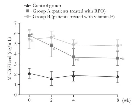
Fig.1.Macrophage-colony stimulating factor(M-CSF)in HCV-related cirrhotic patients treated with RPO supplementation.*:P<0.001,vs healthy controls;#:P<0.05,vs group B.
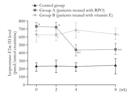
Fig.2.Urinary isoprostane-F2α-III in HCV-related cirrhotic patients:effect of RPO supplementation.*:P<0.05,vs healthy controls;#:P<0.05,vs group B.
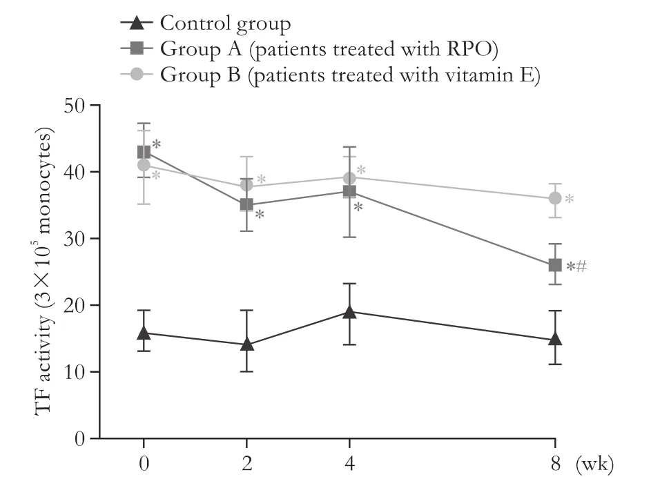
Fig.3.Tissue factor(TF)activity in HCV-related cirrhotic patients:effect of RPO supplementation.*:P<0.05,vs healthy controls;#:P<0.05,vs group B.
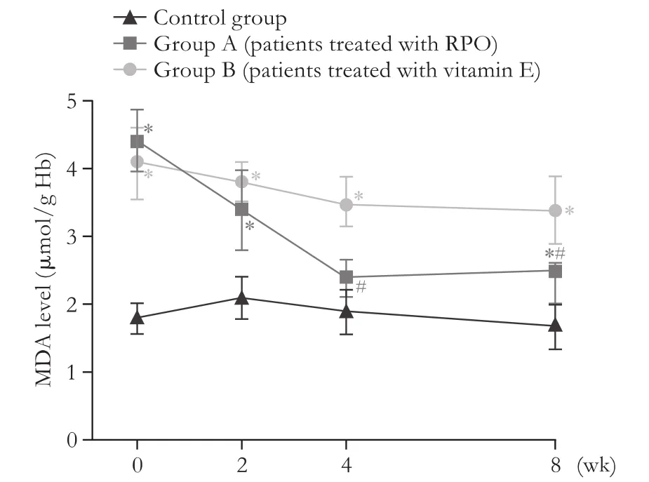
Fig.4.Erythrocyte MDA level in HCV-related cirrhotic patients:effect of RPO supplementation.*:P<0.05,vs healthy controls;#:P<0.05,vs group B.
Assessment of monocyte TF activity
Patients with liver cirrhosis showed a higher mean value of this parameter(Fig.3,P<0.05).It also appeared as a trend of positive direct correlation with M-CSF values(r=0.45,P=0.08).This parameter was unaffected by vitamin E treatment whereas daily RPO supplementation brought about a gradual decrease of this parameterwhich reached a statistically significant change at a 8-week observation(P<0.05).At this stage the parameter and M-CSF values showed a significant correlation(r=0.67,P<0.05).
Measurement of MDA in erythrocytes
As compared to the controls,the erythrocyte level of MDA was significantly higher in cirrhotic patients(P<0.01)but it was beneficially affected by RPO treatment(P<0.05 vs baseline,Fig.4)and at lower,not to a significant extent,by vitamin E supplementation(P>0.05).
Discussion
Oxidative stress is an essential factor for the progression of chronic liver diseases,such as chronic hepatitis C,non-alcoholic steatohepatitis,drug-induced liver injury,and genetic metal storage disorders.
Vitamin E is a fat-soluble vitamin with antioxidant properties.It is known that daily oral vitamin E administration is able to normalize serum aminotransferase and alkaline phosphatase levels,and to significantly improve lobular inflammation.Also RPO,which acts as a potent antioxidant,is linked by several studies to better recovery and protection of oxygen-susceptible cells when exposed to oxidative stress.
Endotoxin represents a fundamental LPS fraction of Gram-negative bacterial cell wall.The release of endotoxin within the intestinal lumen and its further clearance by the liver is regarded as a physiological phenomenon.[28]Thus,the transient presence of endotoxin within the portal system but not in the supra-hepatic vein is a common finding[3]whereas in the case of hepatic insufficiency there is a more substantial spill-over of endotoxin through the liver into the systemic circulation.[29]Our present study demonstrates that a protracted treatment with either vitamin E or RPO did not significantly affect the endotoxin level and endotoxin-inhibiting capacity of plasma,as far as albumin and high density lipoprotein fractions are regarded.Moreover,this variable consists of a rather complex interplay between a number of other parameters such as leukopenia,opsonizing capacity,etc.Hyperendotoxinemia in the course of liver disease further contributes to liver damage by promoting mononuclear cells infiltration,[8,11,30]and it has been recently demonstrated that it can also induce the expression of a chemoattractant protein.[31]Once recruited,such activated monocytes can release a number of cytokines[32,33]which,on their turn,elicit the patho-mechanisms of hepatotoxicity.[34]Accordingly,Itoh et al[35]has proven that in the course of acute and chronic liver failure there is an increase of serum concentration of M-CSF which is a cytokine mostly secreted by monocytes/macrophages endowed by potent chemoattractant properties on monocytes,as very recently confirmed.[36]Further,the same group has showed that serum M-CSF is closely related with proinflammatory cytokines produced by peripheral blood cells during chronic hepatitis.[37]
This study aimed to obtain preliminary results on the effectiveness of RPO therapy in patients with HCV-related cirrhosis.We recognized that the study enrolled a small sample size due to the limitations caused by the numerous exclusion criteria.In our study,did not take into account the possible improvement of liver fibrosis in patients with HCV-related cirrhosis treated with RPO.Fibroscan analyses performed during the study did not show any significant change,probably because of the short duration of the study.
Our study confirmed elevated M-CSF in the cirrhotic patients,which was almost two-fold higher than in the controls.Of interest,supplementation with RPO,probably via the attenuation of endotoxin-mediated stimulation of monocytes,brought about a significant decrease of M-CSF synthesis from macrophage with a reduced serum level of this cytokine and this effect was most significant in RPO-treated patients.The same phenomenon,although a little bit delayed,occurred with TF activity which highly correlated with the RPO-related changes of M-CSF at the end of the study period.However,further studies are needed to better define the transcriptional or post-transcriptional activation of monocyte TF expression.We can envisage that the high content of carotenoids in RPO by regulating intracellular events is likely to be involved in a number of key immune function assays such as immunoglobulin production,lymphocyte cytotoxic activity,and modulating cytokine production.Moreover,such on-going status of pre-activation of polymorphonuclear cells and monocytes/macrophages is negatively influenced by oxidative stress which is usually observed in liver patients[11,38]and which clearly appeared also in our study.We can tentatively suggest that this is the result of the constant exposure of monocytes to high antigenic load,although factors such as unbalanced antioxidant status,depleted intracellular glutathione concentration and others might play a relevant role.Therefore,RPO might have also acted by partially putting monocytes “at rest”,thus enabling a recovery of the inner metabolic oxidative mechanisms.These data may be corroborated by the significant decrease of erythrocyte level of MDA obtained only in RPO-treated patients.Although no difference in radical scavenging activity between α-tocopherol and α-tocotrienol has been found in hexane,the activity of α-tocotrienol in scavenging peroxyl radicals has been shown to be 1.5-foldhigher in liposomes than that of α-tocopherol.[39]It is interesting to consider the richness of both fractions in RPO.Our finding is in agreement with Devaraj and Jialal[40]who suggested that,the negative results yielded by α-tocopherol in a number of prospective clinical trials of dietary antioxidants may be at least partially explained by its decreasing effect of γ-tocopherol levels which has also potent anti-inflammatory activity.Indeed,plasma γ-tocopherol levels are inversely associated with cardiovascular diseases.Unlike allopurinol which has been shown by Spahr et al[41]to significantly decrease oxidative stress in cirrhotic patients but not inflammatory parameters,RPO supplementation without affecting endotoxemia provides a significant down-regulation of related inflammatory molecules.
Although the ultimate therapeutic target is to eradicate HCV,antioxidant-anti-inflammatory nutritional modulation might offer a worthwhile complementary tool.Indeed,the liver is one of the most susceptible organs to oxidative-related cellular damage and DNA mutagenesis,and oxidative stress has been implicated as a causative factor in alcoholic and non-alcoholic liver disease.
In this setting,given the long-term management of such patients,a nutritionally-enriched dietary regimen would represent an ideal approach.In fact,despite a measurable level of saturated fatty acids,the presence of carotenoids and the whole array of tocopherols and tocotrienols which are endowed by powerful anti-oxidants effects make RPO as one of the most advisable vegetable oils with health promoting properties.
However,such complementary support still deserves further studies.Uncontrolled vitamin supplementation has to be discouraged in consideration of the recent review questioning the benefit of high-dosage of synthetic vitamin E[42]as well as the recent experimental data on potential liver toxicity of high-dosage of synthetic delta-alpha-tocopherol supplementation[43]or the reported unexpectedly HCV viremia-enhancing effect of resveratrol.[16]
Acknowledgement:We thank Mr.O.Licciardello from Science of Living,Milano,Italy,for his logistic support.
Contributors:CR and IA enrolled the patients and performed patients’physical examination,upper gastrointestinal endoscopy,testing of blood samples and liver ultrasound.SU,MM and MF designed the study;in addition,MF contributed to overall coordination and,with MM,to correspondence with other authors,while SU contributed to methodology assessments.MD made preliminary in vitro testing and statistical analysis.AA conducted biochemical evaluation and methodology assessments.ZN helped doing preliminary patients recruitment for an observational pilot study testing palatability and compliance feedback together with first biochemical test battery.CR is the guarantor.
Funding:This study was supported by a generous and unbiased grant from the Malaysian Palm Oil Board,Bandar Baru Bangi,43000 Kajang,Selangor,Malaysia.
Ethical approval:This study was approved by the Ethics Committee of A.O.U.“Policlinico-Vittorio Emanuele”,Università di Catania,Italy.
Competing interest:No benefits in any form have been received or will be received from a commercial party related directly or indirectly to the subject of this article.
References
1 Gaeta GB,Perna P,Adinolfi LE,Utili R,Ruggiero G.Endotoxemia in a series of 104 patients with chronic liver diseases:prevalence and significance.Digestion 1982;23:239-244.
2 Tarao K,So K,Moroi T,Ikeuchi T,Suyama T.Detection of endotoxin in plasma and ascitic fluid of patients with cirrhosis:its clinical significance.Gastroenterology 1977;73:539-542.
3 Lumsden AB,Henderson JM,Kutner MH.Endotoxin levels measured by a chromogenic assay in portal,hepatic and peripheral venous blood in patients with cirrhosis.Hepatology 1988;8:232-236.
4 Bengmark S.Bio-ecological control of chronic liver disease and encephalopathy.Metab Brain Dis 2009;24:223-236.
5 Kuhla A,Norden J,Abshagen K,Menger MD,Vollmar B.RAGE blockade and hepatic microcirculation in experimental endotoxaemic liver failure.Br J Surg 2013;100:1229-1239.
6 De Minicis S,Rychlicki C,Agostinelli L,Saccomanno S,Candelaresi C,Trozzi L,et al.Dysbiosis contributes to fibrogenesis in the course of chronic liver injury in mice.Hepatology 2014;59:1738-1749.
7 Lee KC,Yang YY,Wang YW,Lee FY,Loong CC,Hou MC,et al.Increased plasma malondialdehyde in patients with viral cirrhosis and its relationships to plasma nitric oxide,endotoxin,and portal pressure.Dig Dis Sci 2010;55:2077-2085.
8 Amer A,Amer ME.Enhanced monocyte tissue factor expression in hepatosplenic schistosomiasis.Blood Coagul Fibrinolysis 2002;13:43-47.
9 Heredia D,Caballería J,Arroyo V,Ravelli G,Rodés J.Lactitol versus lactulose in the treatment of acute portal systemic encephalopathy(PSE).A controlled trial.J Hepatol 1987;4:293-298.
10 Riggio O,Balducci G,Ariosto F,Merli M,Tremiterra S,Ziparo V,et al.Lactitol in the treatment of chronic hepatic encephalopathy--a randomized cross-over comparison with lactulose.Hepatogastroenterology 1990;37:524-527.
11 Jaeschke H.Reactive oxygen and mechanisms of inflammatory liver injury:Present concepts.J Gastroenterol Hepatol 2011;26:173-179.
12 Ramachandran A,Prabhu R,Thomas S,Reddy JB,Pulimood A,Balasubramanian KA.Intestinal mucosal alterations in experimental cirrhosis in the rat:role of oxygen free radicals.Hepatology 2002;35:622-629.
13 Musiek ES,Yin H,Milne GL,Morrow JD.Recent advances in the biochemistry and clinical relevance of the isoprostane pathway.Lipids 2005;40:987-994.
14 Basu S.Isoprostanes:novel bioactive products of lipid peroxidation.Free Radic Res 2004;38:105-122.
15 Devaraj S,Li D,Jialal I.The effects of alpha tocopherol supplementation on monocyte function.Decreased lipid oxidation,interleukin 1 beta secretion,and monocyte adhesion to endothelium.J Clin Invest 1996;98:756-763.
16 Nakamura M,Saito H,Ikeda M,Hokari R,Kato N,Hibi T,et al.An antioxidant resveratrol significantly enhanced replication of hepatitis C virus.World J Gastroenterol 2010;16:184-192.
17 Szucs G,Bester DJ,Kupai K,Csont T,Csonka C,Esterhuyse AJ,et al.Dietary red palm oil supplementation decreases infarct size in cholesterol fed rats.Lipids Health Dis 2011;10:103.
18 Aboua YG,Brooks N,Mahfouz RZ,Agarwal A,du Plessis SS.A red palm oil diet can reduce the effects of oxidative stress on rat spermatozoa.Andrologia 2012;44:32-40.
19 Subar AF,Thompson FE,Kipnis V,Midthune D,Hurwitz P,McNutt S,et al.Comparative validation of the Block,Willett,and National Cancer Institute food frequency questionnaires:the Eating at America’s Table Study.Am J Epidemiol 2001;154:1089-1099.
20 Fukui H,Brauner B,Bode JC,Bode C.Plasma endotoxin concentrations in patients with alcoholic and non-alcoholic liver disease:reevaluation with an improved chromogenic assay.J Hepatol 1991;12:162-169.
21 Bode C,Fukui H,Bode JC.“Hidden” endotoxin in plasma of patients with alcoholic liver disease.Eur J Gastroenterol Hepatol 1993;5:247-262.
22 Fukui H,Brauner B,Bode JC,Bode C.Chromogenic endotoxin assay in plasma.Selection of plasma pretreatment and production of standard curves.J Clin Chem Clin Biochem 1989;27:941-946.
23 Fukui H,Tsujita S,Matsumoto M,Morimura M,Kitano H,Kinoshita K,et al.Endotoxin inactivating action of plasma in patients with liver cirrhosis.Liver 1995;15:104-109.
24 Hanamura T,Motoyoshi K,Yoshida K,Saito M,Miura Y,Kawashima T,et al.Quantitation and identification of human monocytic colony-stimulating factor in human serum by enzyme-linked immunosorbent assay.Blood 1988;72:886-892.
25 Shadle PJ,Allen JI,Geier MD,Koths K.Detection of endogenous macrophage colony-stimulating factor(M-CSF)in human blood.Exp Hematol 1989;17:154-159.
26 Mori TA,Croft KD,Puddey IB,Beilin LJ.An improved method for the measurement of urinary and plasma F2-isoprostanes using gas chromatography-mass spectrometry.Anal Biochem 1999;268:117-125.
27 Jain SK.Membrane lipid peroxidation in erythrocytes of the newborn.Clin Chim Acta 1986;161:301-306.
28 Ruiter DJ,van der Meulen J,Brouwer A,Hummel MJ,Mauw BJ,van der Ploeg JC,et al.Uptake by liver cells of endotoxin following its intravenous injection.Lab Invest 1981;45:38-45.
29 Lin RS,Lee FY,Lee SD,Tsai YT,Lin HC,Lu RH,et al.Endotoxemia in patients with chronic liver diseases:relationship to severity of liver diseases,presence of esophageal varices,and hyperdynamic circulation.J Hepatol 1995;22:165-172.
30 Itoh Y,Okanoue T,Enjo F,Sakamoto S,Keizo K,Yasukazu O,et al.Macrophage colony stimulating factor and hepatitis.Int Hepatol Commun 1993;1:1-4.
31 Czaja MJ,Geerts A,Xu J,Schmiedeberg P,Ju Y.Monocyte chemoattractant protein 1(MCP-1)expression occurs in toxic rat liver injury and human liver disease.J Leukoc Biol 1994 Jan;55(1):120-126.
32 Müller C,Zielinski CC.Interleukin-6 production by peripheral blood monocytes in patients with chronic liver disease and acute viral hepatitis.J Hepatol 1992;15:372-377.
33 Deviere J,Content J,Denys C,Vandenbussche P,Schandene L,Wybran J,et al.High interleukin-6 serum levels and increased production by leucocytes in alcoholic liver cirrhosis.Correlation with IgA serum levels and lymphokines production.Clin Exp Immunol 1989;77:221-225.
34 Itoh Y,Okanoue T,Enjyo F,Sakamoto S,Ohmoto Y,Hirai Y,et al.Serum levels of macrophage colony stimulating factor(MCSF)in liver disease.J Hepatol 1994;21:527-535.
35 Itoh Y,Okanoue T,Sakamoto S,Nishioji K,Kashima K.The effects of prednisolone and interferons on serum macrophage colony stimulating factor concentrations in chronic hepatitis B.J Hepatol 1997;26:244-252.
36 Däbritz J,Bonkowski E,Chalk C,Trapnell BC,Langhorst J,Denson LA,et al.Granulocyte macrophage colony-stimulating factor auto-antibodies and disease relapse in inflammatory bowel disease.Am J Gastroenterol 2013;108:1901-1910.
37 Itoh Y,Okanoue T,Ohnishi N,Sakamoto M,Nishioji K,Nakagawa Y,et al.Serum levels of soluble tumor necrosis factor receptors and effects of interferon therapy in patients with chronic hepatitis C virus infection.Am J Gastroenterol 1999;94:1332-1340.
38 Choi J,Corder NL,Koduru B,Wang Y.Oxidative stress and hepatic Nox proteins in chronic hepatitis C and hepatocellular carcinoma.Free Radic Biol Med 2014;72:267-284.
39 Serbinova E,Kagan V,Han D,Packer L.Free radical recycling and intramembrane mobility in the antioxidant properties of alpha-tocopherol and alpha-tocotrienol.Free Radic Biol Med 1991;10:263-275.
40 Devaraj S,Jialal I.Failure of vitamin E in clinical trials:is gamma-tocopherol the answer? Nutr Rev 2005;63:290-293.
41 Spahr L,Bresson-Hadni S,Amann P,Kern I,Golaz O,Frossard JL,et al.Allopurinol,oxidative stress and intestinal permeability in patients with cirrhosis:an open-label pilot study.Liver Int 2007;27:54-60.
42 Dotan Y,Lichtenberg D,Pinchuk I.No evidence supports vitamin E indiscriminate supplementation.Biofactors 2009;35:469-473.
43 Asare GA,Ntombini B,Kew MC,Kahler-Venter CP,Nortey EN.Possible adverse effect of high delta-alpha-tocopherol intake on hepatic iron overload:enhanced production of vitamin C and the genotoxin,8-hydroxy-2’-deoxyguanosine.Toxicol Mech Methods 2010;20:96-104.
Received January 30,2015
Accepted after revision September 25,2015
doi:10.1016/S1499-3872(16)60072-3
Corresponding Author:Professor Roberto Catanzaro,MD,PhD,Presidio Ospedaliero Policlinico “G.Rodolico”,U.O.C.di Medicina Interna “A.Francaviglia”,Bldg.n.4 - I° Floor,Via S.Sofia,78,95123 Catania,Italy(Tel:+39-95-3782902;Fax:+39-95-3782376;Email:rcatanza@unict.it)
 Hepatobiliary & Pancreatic Diseases International2016年2期
- Hepatobiliary & Pancreatic Diseases International的其它文章
- Hepatobiliary &Pancreatic Diseases International
- Hyaluronic acid algorithm-based models for assessment of Iiver fibrosis:transIation from basic science to clinical application
- Liver transplantation for hepatocellular carcinoma:is zero recurrence theoretically possible?
- Cancer of the Liver Italian Program score helps identify potential candidates for transarterial chemoembolization in patients with Barcelona Clinic Liver Cancer stage C
- Efficient generation of functionaI hepatocyte-Iike ceIIs from mouse Iiver progenitor ceIIs via indirect co-cuIture with immortaIized human hepatic stellate cells
- Individualized nomogram improves diagnostic accuracy of stage I-II gallbladder cancer in chronic cholecystitis patients with gallbladder wall thickening
