两种转移皮瓣成形术治疗骶尾部藏毛窦
作者单位:100120 北京,北京市肛肠医院(二龙路医院)肛肠外科外三病区
两种转移皮瓣成形术治疗骶尾部藏毛窦
于锦利段宏岩王凯蔡姮婧孙磊郝鹏
【摘要】目的评价及比较2种转移皮瓣成形术治疗骶尾部藏毛窦的疗效。方法96例骶尾部藏毛窦患者分别接受改良Limberg皮瓣成形术(Limberg组,28例)和改良Karydakis 皮瓣成形术(Karydakis组,68例)治疗。记录和观察患者的手术时间、术后皮瓣坏死情况、伤口愈合情况、住院时间,术后随访至少3个月,观察复发情况。对2组数据进行比较。结果Limberg 组手术时间较Karydakis组长[中位数(四分位数间距)为110(3.75)min vs. 90(40)min],皮瓣水泡、表皮坏死发生率较Karydakis组高(25% vs.0),伤口一期愈合率低于Karydakis组(46% vs.71%),住院时间长于Karydakis组[中位数(四分位数间距)为20(4.75)d vs. 17(9)d](P均<0.05)。随访3~49(23.5)个月,2组复发率比较差异无统计学意义(P>0.05)。 结论改良Limberg皮瓣成形术及改良Karydakis皮瓣成形术均是治疗骶尾部藏毛窦的有效方法,改良Karydakis皮瓣成形术的疗效更好。
【关键词】藏毛窦;转移皮瓣;手术;疗效
DOI:10.3969/g.issn.0253-9802.2015.03.006
收稿日期:(2014-09-15)
Application of two types of flap transposition plasty in treatment of sacrococcygeal pilonidal sinusYuJinli,DuanHongyan,WangKai,CaiHengjing,SunLei,HaoPeng.DepartmentofAnorectalSurgery,Ward3,BeijingAnorectalHospital(BeijingErlongRoadHospital),Beijing100120,China
Abstract【】ObjectiveTo compare the clinical efficacy of two types of flap transposition plasty in the treatment of sacrococcygeal pilonidal sinus. MethodsA total of 96 patients with sacrococcygeal pilonidal sinus were divided into the modified Limberg flap plasty (Limberg group, n=28) and modified Karydakis flap plasty groups (Karydakis group, n=68). The operation time, postoperative flap necrosis and wound healing, length of hospital stay were observed and recorded. Postoperative follow-up endured for ≥ 3 months. The recurrence rate of sacrococcygeal pilonidal sinus was observed. All these parameters were statistically compared between two groups. ResultsIn the Limberg group, the operation time(median: interquartile range) was 110(3.75) min, significantly longer than 90(40) min in the Karydakis group. The incidence of flap blister and epidermal necrosis was 25%, significantly higher compared with 0% in the Karydakis group. The percentage of patients with stage I wound healing was 46%, significantly less than 71% in the Karydakis group, and the length of hospital stay(median: interquartile range) was 20 (4.75) d which was significantly longer compared with 17 (9) d in the Karydakis group (all P<0.05). Time of follow-up ranged from 3 to 49 months (23.5 month on average). The recurrence rate did not significantly differ between two groups (P>0.05). ConclusionsAlbeit both modified Limberg and Karydakis flap plasty are efficacious treatment of sacrococcygeal pilonidal sinus, the clinical efficacy of the modified Karydakis flap plasty yields better clinical efficacy compared with the modified Limberg flap plasty.
【Key words】Pilonidal sinus; Flap transposition; Surgery; Efficacy
骶尾部藏毛疾病是原发于臀沟并向上蔓延的骶尾部慢性皮下感染, 常反复破溃而形成窦道即藏毛窦。本病好发于青春期,多见于肥胖、毛发厚重者。国内多采用切开引流术,易造成此病的反复复发,国外多采用转移皮瓣成形术,并推荐其为复发性藏毛窦或病灶广泛者的首选治疗方法。我院收治的骶尾部藏毛窦患者多为外院手术治疗失败或复发者,如果切除病灶后敞开创口, 则愈合时间会很长,我们采用了2种改良转移皮瓣成形术治疗本病患者, 现将结果报告如下,以供临床参考。
对象与方法
一、研究对象
2010 年9月至2014年7月我院采用改良转移皮瓣成形术治疗骶尾部藏毛窦患者共96例,其中58 例为外院手术后复发者,24例曾行2次以上手术。所有患者均符合2009或2011年版美国结直肠外科医师协会(ASCRS)指南中的骶尾部藏毛窦诊断标准,即均有骶尾部反复破溃病史及中线处皮肤小凹,术前均行骶尾部MRI确定病变范围及深度, 同时排除骶尾骨疾病。按就诊顺序,于2010年9月至2011年10月就诊的28例患者接受改良Limberg 皮瓣成形术治疗(Limberg组),于2011年11月至2014年7月就诊的68例患者接受改良Karydakis皮瓣成形术治疗(Karydakis组)。Limberg组男27例、女1例,年龄14~37岁、中位年龄23岁,中线处皮肤小凹距骶尾部破溃处3~7 cm、中位数4 cm;Karydakis组男58例、女10例,年龄15~49岁、中位年龄23岁,中线处皮肤小凹距骶尾部破溃处3~8 cm、中位数4 cm,2组性别、年龄等比较差异无统计学意义(P均>0.05)。
二、治疗
1.术前准备
术前,臀沟、骶尾部手术区常规备皮。手术前1 d口服复方聚乙二醇4 000口服溶液用粉(4袋,将袋内散剂溶于2 000 ml水中服用),彻底排空肠道。手术当日早晨禁水、禁食。设计、标记切除区与皮瓣区。
2.手术方法
采取连续性硬膜外阻滞或全身麻醉, 麻醉后患者取俯卧位, 两腿轻微外展, 臀部用胶带左右分开。完整切除包括窦道在内的所有受累组织及中线处皮肤小凹,保留正常皮下组织, 电灼止血,冲洗伤口。按照预设计标记线,游离改良Limberg 皮瓣或改良Karydakis 皮瓣。Limberg组采用改良Limberg皮瓣即菱形皮瓣,根据病变位置确定菱形切除范围,底角需偏离臀沟中线2 cm,切除范围包括骶尾部病灶及中线处皮肤小凹,切除深度为只需将病变整块切除的深度,无需深达骨膜[1]。Karydakis组根据病变位置在偏离臀沟中线一侧2 cm行椭圆形切口,切除范围包块骶尾部病灶及中线处皮肤小凹,无需切除正常组织至骨膜,在对侧切缘游离改良Karydakis皮瓣[2-3]。将皮瓣无张力地覆盖缺损,逐层缝合切口,于皮瓣下放置负压引流管, 用弹力腹带加压包扎伤口。切除标本送病理检查。
3.术后处理
术后,要求患者采用侧卧或俯卧位休息,避免平躺压迫伤口。患者口服蛋白粉,控制饮食,1周内避免下床活动及排大便。用广谱抗生素6 d预防感染。每日换药时观察伤口生长情况,臀沟处用纱布垫高,用弹力腹带加压包扎伤口。术后2~3 d若引流无新鲜血液且全日引流量少于5 ml则可拔除引流管。术后10~14 d拆线。出院后每周回院复查,随访至少3个月,指导患者脱毛,范围由肛门后方经臀沟至后背平坦处,伤口未完全愈合时用小镊子拔除伤口周围毛发,防止毛发落入伤口,伤口完全愈合后采用脱毛膏或行激光脱毛。同时观察骶尾部藏毛窦有否复发。
三、观察指标
记录和观察患者的手术时间、术后皮瓣坏死情况、伤口愈合情况、住院时间,术后随访至少3个月,观察复发情况。对2组数据进行比较。
四、统计学处理
采用SPSS 19.0 软件处理数据,计量资料用中位数(四分位数间距)表示,组间比较用秩和检验。计数资料组间比较采用χ2检验或Fisher确切概率法。P<0.05为差异具有统计学意义。
结果
一、一般情况
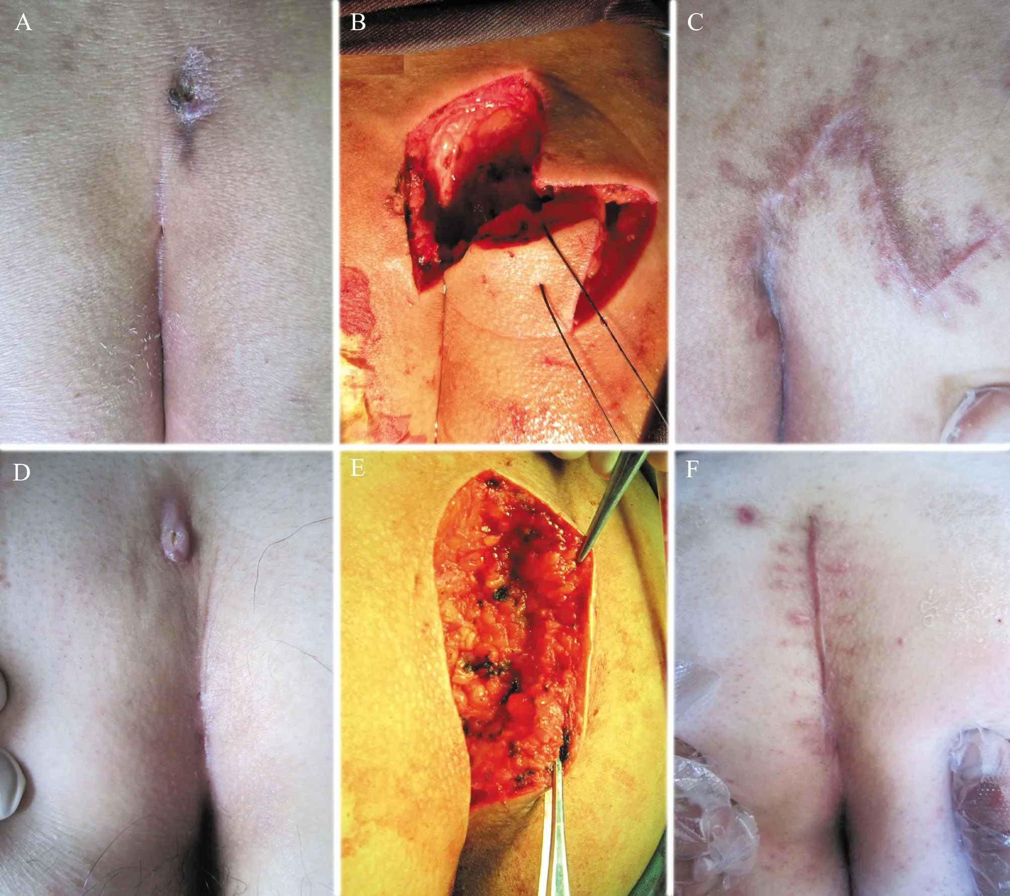
96例患者术程均顺利,出血量少,术后61例(64%)伤口一期愈合,住院时间15~23(18)d。Limberg组图例见图1A~C,Karydakis组图例见图1D~F。Limberg组15例术后伤口部分裂开,经换药后二期愈合,1例术后皮瓣下方出现血肿需行二次手术止血。Karydakis组20例术后伤口部分裂开,经换药后二期愈合;1例术后伤口出现化脓性感染需行二次手术切开引流。
二、Limberg组与Karydakis组各观察指标的比较
Limberg组手术时间较Karydakis组长,皮瓣水泡、表皮坏死发生率较Karydakis组高,伤口一期愈合率低于Karydakis组,住院时间长于Karydakis组(P均<0.05),见表1。
三、随访情况
所有患者随访3~49(中位数23.5)个月,Limberg组1例(3.57%)术后3年伤口下方肿胀复发,再次接受手术切开治疗;Karydakis组1例(1.47%)术后1个月伤口下方肿胀复发,再次接受手术切开治疗,2组复发率比较差异无统计学意义(P=0.500)。其余患者随访情况良好。
表1

Limberg 组与Karydakis组各观察指标的比较

图1 Limberg组及Karydakis组转移皮瓣成形术手术图例
A~C:Limberg组术前臀沟病灶、术中切除区、术后皮瓣转移处恢复情况;D~F: Karydakis组术前臀沟病灶、术中切除区、术后皮瓣转移处恢复情况
讨论
藏毛窦的形成有先天性和后天性2种学说。Karydakis[2]认为骶尾部藏毛窦是由臀沟处毛发刺入引起的,且松散的毛发可来自身体其他部位。松散的毛发、臀沟摩擦产生的力和臀中线皮肤的易损性是毛发刺入的3个主要因素。Bascom等[3]认为臀沟中线处的小凹为增大扩张的毛囊,毛发由此进入,潮湿、缺氧、毛发和细菌造成的表皮和深部组织破坏加速藏毛窦的形成。目前,藏毛窦的后天性形成学说已逐渐被大家所接受,检查患者可发现其臀沟中线的小凹,即原发口,本研究中96例患者均存在中线处皮肤小凹。
一、骶尾部藏毛窦切除缝合的手术原则
为了改善臀沟处皮肤的易损性,避免毛发刺入,Karydakis和Bascom分别设计了采用皮瓣覆盖创面的手术方法,有效地降低了患者的复发率,其后,此2种手术方法被多位学者采用并进行了改良,取得了良好的疗效[2-6]。2种手术方法的共同原则为:①完全切除中线处皮肤小凹;②通过皮瓣抬高臀沟;③避免出现中线缝合伤口;④减少缝合张力,促进伤口愈合。2种手术方法不同之处是,Karydakis完整切除病灶,深达骨膜,而Bascom认为不需要切除深部组织,而且不必切除继发病灶,仅切除中线处皮肤小凹、游离皮肤皮瓣覆盖创面即可。有学者报道,Limberg皮瓣成形术及其改良术式亦能达到完全切除中线处皮肤小凹、抬高臀沟、避免出现中线缝合伤口的目的,而且能减少张力、提高一期愈合率、缩短愈合时间[7-16]。我们发现,固定皮瓣角度及切除病灶深达骶骨筋膜并不适合所有患者,因此我们根据患者病灶情况适当调整皮瓣角度及切除深度,采用改良Limberg 皮瓣成形术和改良Karydakis皮瓣成形术治疗骶尾部藏毛窦,术后64%患者的伤口一期愈合,提示转移皮瓣成形术是治疗骶尾部藏毛窦的有效方法。
二、2种皮瓣的比较
改良Limberg皮瓣为旋转皮瓣,需增加额外切口,为了防止旋转成角,缓解张力,需切除及游离更多的皮下组织。改良Karydakis皮瓣仅游离病灶对侧的切缘皮肤,无需增加额外切口[3]。从我们的研究可以看出,2组术后复发率比较差异无统计学意义,Karydakis组疗效更好。我们分析Limberg组术后皮瓣水泡、表皮坏死发生率较高的原因可能有:①改良Limberg皮瓣游离组织范围大,切口长;②缝合改良Limberg皮瓣后会形成伤口侧方及纵向张力,侧方张力可通过弹力腹带缓解,但纵向张力不能完全缓解;③改良Limberg皮瓣缝合造成伤口多处成角,不利于伤口对合。有研究结果显示,改良Karydakis皮瓣成形术与改良Limberg皮瓣成形术术后复发率比较无差异,但Karydakis皮瓣成形术后并发症发生率、术后疼痛程度、手术时间均优于Limberg皮瓣成形术,患者满意度更高[17-20]。我们的研究结果也显示Karydakis皮瓣成形术疗效更好。然而也有研究结果显示改良Limberg皮瓣成形术较改良Karydakis皮瓣成形术的疗效好[21-23]。有学者的的系统综述及荟萃分析显示中线缝合术后伤口感染及裂开发生率明显高于偏离中线缝合,但比较Karydakis皮瓣与Limberg 皮瓣,两者术后复发率及伤口并发症发生率无明显差异[24-26]。因此,选择何种皮瓣才能达到最佳治疗效果,仍应进行更大样本的随机对照研究来证实。
三、围手术期处理
适当的围手术期处理对避免出现术后并发症有重要意义。术前排空肠道,术后控制排大便及避免活动可有效减少伤口污染及切口移位,我们观察到,转移皮瓣成形术后臀沟处伤口愈合欠佳多与患者过早排大便及活动有关,因此,我们建议患者术后口服蛋白粉,控制饮食,1周内尽量避免下床活动及排大便,但仍有部分患者依从性较差。有学者提出,Karydakis皮瓣成形术后可不放置引流管[27]。我们观察到1例患者引流管过早脱落后出现皮瓣下方血肿,需再次接受手术治疗,因此我们建议常规放置负压引流管[1]。术后患者应采用侧卧或俯卧位,避免平躺,用弹力腹带加压包扎伤口可有效缓解伤口张力,这些做法均有助于避免切口周围产生张力性水泡继而造成表皮坏死,本研究中Limberg组有7例出现皮瓣水泡、表皮坏死,考虑与在年份较早时进行的手术没注意叮嘱患者术后采用侧卧或俯卧位及采用弹力腹带加压包扎有关。
四、总结
改良Limberg皮瓣成形术及改良Karydakis皮瓣成形术均是治疗骶尾部藏毛窦的有效方法,改良Karydakis皮瓣手术简单、疗效更好,但仍有部分患者术后会出现伤口裂开,因此,如何提高伤口一期愈合率还有待我们行更大样本的研究。
参考文献
[1]段宏岩, 刘连成, 于锦利,等. 改良Limberg、Dufourmentel菱形转移皮瓣成形术治疗骶尾部藏毛窦. 中华整形外科杂志, 2012, 28: 69-71.
[2]Karydakis GE. New approach to the problem of pilonidal sinus. Lancet, 1973, 2: 1414-1415.
[3]Bascom J, Bascom T. Failed pilonidal surgery: new paradigm and new operation leading to cures. Arch Surg, 2002, 137: 1146-1150.
[4]Iesalnieks I, Deimel S, Schlitt HJ. Karydakis flap for recurrent pilonidal disease. World J Surg, 2013, 37: 1115-1120.
[5]Guner A, Ozkan OF, Kece C, et al. Modification of the Bascom cleft lift procedure for chronic pilonidal sinus: results in 141 patients. Colorectal Dis, 2013, 15: 402-406.
[6]Yildiz MK, Ozkan E, Odabai HM, et al. Karydakis flap procedure in patients with sacrococcygeal pilonidal sinus disease: experience of a single centre in Istanbul. Scientific World Journal, 2013, 2013: 807027.
[7]董明君, 周宝峰, 戴晓宇. 窦道切除加菱形皮瓣转移治疗骶尾部藏毛窦临床分析. 现代实用医学, 2012, 24: 315-316.
[8]黄士勇, 王中川, 孙基伟, 等. 菱形皮瓣转移缝合术治疗复发的骶尾部藏毛窦. 中华临床医师杂志:电子版, 2012, 6: 8383-8384.
[9]Kaya B, Eris C, Atalay S, et al. Modified Limberg transposition flap in the treatment of pilonidal sinus disease.Tech Coloproctol, 2012, 16: 55-59.
[10]Karakas BR. Comparison of Z-plasty, Limberg flap, and asymmetric modified Limberg flap techniques for the pilonidal sinus treatment: review of literature. Acta Chir Iugosl, 2013, 60: 31-37.
[11]Aithal SK, Rajan CS, Reddy N. Limberg flap for sacrococcygeal pilonidal sinus a safe and sound procedure. Indian J Surg, 2013, 75: 298-301.
[12]Yabanoglu H, Karagulle E, Belli S, et al. Results of modified Dufourmentel rhomboid flap in patients with extensive sacrococcygeal pilonidal disease. Acta Chir Belg, 2014, 114: 52-57.
[13]Dass TA, Zaz M, Rather A, et al. Elliptical excision with midline primary closure versus rhomboid excision with limberg flap reconstruction in sacrococcygeal pilonidal disease: a prospective, randomized study. Indian J Surg, 2012, 74: 305-308.
[14]Karaca AS, Ali R, Capar M, et al. Comparison of Limberg flap and excision and primary closure of pilonidal sinus disease, in terms of quality of life and complications. J Korean Surg Soc, 2013, 85: 236-239.
[15]Khan PS, Hayat H, Hayat G. Limberg flap versus primary closure in the treatment of primary sacrococcygeal pilonidal disease; a randomized clinical trial. Indian J Surg, 2013, 75: 192-194.
[16]Duman K, Ozdemir Y, Yucel E, et al. Comparison of depression, anxiety and long-term quality of health in patients with a history of either primary closure or Limberg flap reconstruction for pilonidal sinus. Clinics (Sao Paulo), 2014, 69: 384-387.
[17]Ates M, Dirican A, Sarac M, et al. Short and long-term results of the Karydakis flap versus the Limberg flap for treating pilonidal sinus disease: a prospective randomized study. Am J Surg, 2011, 202: 568-573.
[18]Bessa SS. Comparison of short-term results between the modified Karydakis flap and the modified Limberg flap in the management of pilonidal sinus disease: a randomized controlled study. Dis Colon Rectum, 2013, 56: 491-498.
[19]Guner A, Boz A, Ozkan OF,et al. Limberg flap versus Bascom cleft lift techniques for sacrococcygeal pilonidal sinus:prospective, randomized trial. World J Surg, 2013, 37: 2074-2080.
[20]Orhalmi J, Sotona O, Dušek T,et al. Pilonidal sinus - possibilities surgical treatment. Rozhl Chir, 2014, 93: 491-495.
[21]Arslan K, Said Kokcam S, Koksal H, et al. Which flap method should be preferred for the treatment of pilonidal sinus? A prospective randomized study. Tech Coloproctol, 2014, 18: 29-37.
[22]Sit M, Aktas G, Yilmaz EE. Comparison of the three surgical flap techniques in pilonidal sinus surgery.Am Surg, 2013, 79: 1263-1268.
[23]Karaca T, Yoldaʂ O, Bilgin BÇ, et al. Comparison of short-term results of modified Karydakis flap and modified Limberg flap for pilonidal sinus surgery. Int J Surg, 2012, 10: 601-606.
[24]Horwood J, Hanratty D, Chandran P, et al. Primary closure or rhomboid excision and Limberg flap for the management of primary sacrococcygeal pilonidal disease? A meta-analysis of randomized controlled trials. Colorectal Dis, 2012, 14: 143-151.
[25]Enriquez-Navascues JM, Emparanza J, Alkorta M, et al. Meta-analysis of randomized controlled trials comparing different techniques with primaryclosure for chronic pilonidal sinus.Tech Coloproctol, 2014, 18: 863-872.
[26]侯孝涛, 邵万金, 陈玉根. 藏毛窦转移皮瓣技术研究进展.中华普通外科杂志, 2014, 29: 406-408.
[27]Milone M, Di Minno MN, Musella M, et al. The role of drainage after excision and primary closure of pilonidal sinus: a meta-analysis.Tech Coloproctol, 2013, 17: 625-630.
(本文编辑:洪悦民)
临床研究论著

