国产外周全血γ干扰素实验对肺结核病的诊断价值
闫 鹏,任飞霏,苏龙翔,肖 坤,贾艳红,赵志锐,陈 勇,张有江,解立新解放军总医院 呼吸科,北京 0085;协和医院 重症医学科,北京 000;解放军总医院海南分院 呼吸科,海南三亚 570;解放军总院 微生物科,北京 0085
国产外周全血γ干扰素实验对肺结核病的诊断价值
闫 鹏1,任飞霏1,苏龙翔2,肖 坤1,贾艳红3,赵志锐1,陈 勇3,张有江4,解立新1
1解放军总医院 呼吸科,北京 100853;2协和医院 重症医学科,北京 100140;3解放军总医院海南分院 呼吸科,海南三亚 572013;4解放军总院 微生物科,北京 100853
目的探讨国产酶联免疫吸附试验法检测结核特异性细胞免疫反应(简称A.TB检测)对肺结核病的诊断价值。方法2013年5 - 11月在解放军总医院海南分院就诊的疑似肺结核患者326例,均进行A.TB检测,排除资料不全和最终不能确诊病例,共318例纳入分析,其中确诊肺结核患者89例,回顾性分析A.TB对于肺结核病诊断的价值。结果在确诊为肺结核的89例患者中,涂阳28例,其中经A.TB检测阳性20例,阳性率为71.4%;涂阴61例,其中经A.TB检测阳性45例,阳性率为73.8%。对于确诊为肺结核中的涂阳患者、涂阴患者及非结核患者,A.TB的浓度呈明显降低趋势,且差异有统计学意义(χ2=97.360,P=0.001)。涂阳与涂阴患者的A.TB浓度水平均明显高于非结核患者(P=0.001)。入组患者A.TB浓度的ROC曲线下面积为0.816(P=0.001),敏感性为71.9%,特异性为86.1%;涂阳患者A.TB浓度的ROC曲线下面积为0.762(P=0.001),敏感性为71.4%,特异性为87.8%;涂阴患者A.TB浓度的ROC曲线下面积为0.841(P=0.001),敏感性为72.1%,特异性为86.5%。结论国产A.TB可以作为肺结核病的辅助诊断手段。
肺结核;γ干扰素释放试验;临床实验室技术
结核病是严重威胁人类健康的传染病之一。近年来,结核病的发病率有上升趋势。结核菌可侵犯全身各个系统,导致临床表现复杂多变,尤其是肺外结核常因病情隐匿或表现不典型,极易误诊、漏诊[1]。临床用于诊断结核的方法有限,因此发展快速而敏感的实验室诊断技术十分重要。目前国内外常用的是γ干扰素释放试验(interferon gamma release assay,IGRA)。该实验有两种不同检测方法,即酶联免疫吸附试验和酶联免疫斑点试验。酶联免疫吸附试验法即检测结核特异性细胞免疫反应(简称A.TB检测),为一种新的γ干扰素释放试验的方法,由于其经济、快捷、方便的特点,近几年在国内临床诊断中被越来越多地使用。本文拟探讨国产A.TB对肺结核病患者的诊断价值,为临床医生诊断结核提供参考依据。
对象和方法
1 研究对象 2013年5 - 11月在解放军总医院海南分院就诊的临床怀疑肺结核患者326例,行A.TB检查,排除资料不全或最终不能确诊病例,共纳入分析318例检测A.TB的患者。
2 肺结核诊断标准 1)涂阳:标本中涂片可见分枝杆菌或培养为阳性的细菌,鉴定后为结核分枝杆菌;2)涂阴:3次痰涂片及1次痰培养阴性的肺结核,其诊断标准为:①典型肺结核临床症状和胸部X线表现;②抗结核治疗有效;③临床可排除其他非结核性肺部疾病;④结核菌素纯蛋白衍生物(purified protein derivative tuberculin,PPD)(5TU)强阳性;血清抗结核抗体阳性;⑤痰结核菌PCR+探针检测阳性;⑥肺外组织病理证实结核病变;⑦肺泡灌洗液检出抗酸分枝杆菌;⑧支气管或肺组织病理学证实结核病变;具备①~⑥中的3项或⑦~⑧中任何1项可确诊。
3 分组 纳入分析的318例患者,确诊为肺结核病患者(包括涂阳、涂阳患者)为结核组,确诊为非结核病患者为非结核组。
4 样本采集机实验室检测 入选病例均采集5 ml肝素钠抗凝的静脉全血,室温保存。应用结核特异性细胞免疫反应监测(ELISA法,简称A.TB检测)试剂盒(海口维瑅瑗生物研究院产品,批号:201011001)检测A.TB浓度。检测流程:采血16 h内分别用阴性刺激、阳性刺激和抗原刺激,37℃22~24 h后收集血浆,检测γ干扰素含量。结果判定:参照A.TB试剂盒说明书,当阴性刺激剂<500 IU/L、抗原-阴性刺激剂/阳性刺激剂-阴性刺激剂≥0.60时或阴性刺激剂≥500 IU/L、抗原-阴性刺激剂/阳性刺激剂-阴性刺激剂≥0.85时,均判定为阳牲,否则为阴性。
5 统计学处理 采用SPSS18.0统计软件进行统计学分析,计量资料采用±s表示,两组之间比较采用χ2分析。对于非正态或方差不齐的数据,使用中位数(四分位数间距)描述,并应用秩和检验。计数资料用率表示,组间比较采用χ2检验。利用受试者工作特征曲线(ROC曲线),计算A.TB对肺结核的诊断效能和曲线下面积,P<0.05为差异有统计学意义。
结 果
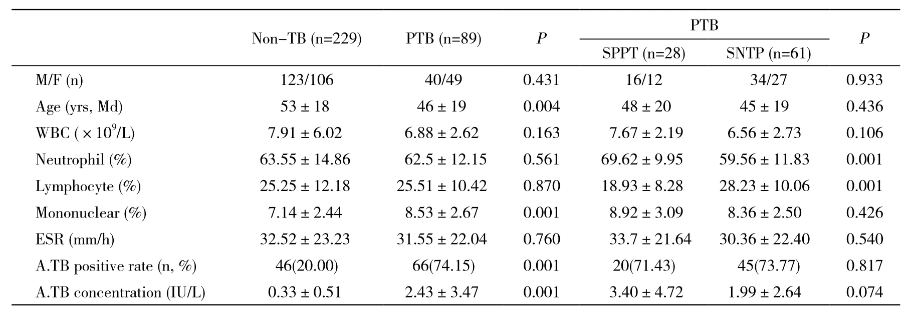
1 一般情况 在纳入分析的318例中,89例为确诊肺结核病例。结核组中涂阳患者28例,A.TB浓度检测阳性20例,阳性率为71.4%;涂阴患者61例,A.TB浓度检测阳性45例,阳性率为73.8%。结核组与非结核组比较,单核细胞计数和A.TB浓度在结核组中均数更大,且两组差异有统计学意义(P=0.001,P=0.001),A.TB浓度的阳性率比较,差异有统计学意义(χ2=82.126,P=0.001);结核组中,涂阳患者和涂阴患者比较,A.TB浓度差异无统计学意义(P=0.074),A.TB浓度的阳性率比较,差异无统计学意义(χ2=0.053,P=0.817)。见表1。
2 不同分组之间A.TB的浓度比较 涂阳患者、涂阴患者、非结核患者A.TB浓度呈明显降低趋势,且差异有统计学意义(χ2=97.360,P=0.001)。3组之间两两比较发现,涂阳与涂阴患者的A.TB浓度均明显高于非结核组患者(P=0.001)。尽管涂阳患者的A.TB浓度高于涂阴患者,但是两者无统计学差异(P=0.839)。见图1。

图 1 不同分组之间A.TB浓度比较Fig. 1 Comparison of A.TB concentration between different groups
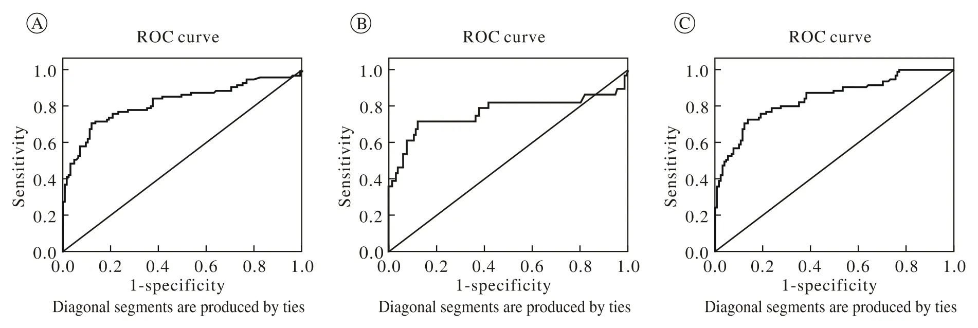
3 A.TB对于肺结核诊断的准确率 所有患者A.TB浓度的ROC曲线下面积为0.816(P=0.001),敏感性为71.9%,特异性为86.1%,准确率为78.31%;涂阳患者A.TB浓度的ROC曲线下面积为0.762(P= 0.001),敏感性为71.4%,特异性为87.8%,准确率为78.99%;涂阴患者A.TB浓度的ROC曲线下面积为0.841(P=0.001),敏感性为72.1%,特异性为86.5%,准确率为78.63%。即A.TB对涂阳结核患者的特异性最好,准确率最高,对涂阴患者诊断的敏感性最好。A.TB浓度对于肺结核诊断的阳性预测值为58.93%,阴性预测值为88.83%,对于涂阴患者阴性预测值91.96%,远大于阳性预测值。A.TB浓度的阳性似然比>5,阴性似然比<1,说明A.TB浓度的高低对于结核诊断有增强作用。见表2和图2。

表1 疑似肺结核患者的基本资料Tab. 1 Basic information of suspected pulmonary tuberculosis patients

表2 A.TB浓度对于不同分组的敏感性、特异性的比较Tab. 2 Comparison of A.TB concentration on the sensitivity and specificity of different groups

图 2 应用ELISA方法,A.TB(ELISA法)对肺结核诊断效率的ROC曲线 A: PTB; B: SPPT; C: SNPTFig. 2 ROC curve of A.TB in patients with suspected tuberculosis by ELISA A: PTB; B: SPPT; C: SNPT
讨 论
临床中诊断结核存在很大的困难,传统的结核诊断方法敏感性和特异性都很差[2-3]。痰涂片和痰培养寻找结核分枝杆菌敏感性极差,但具有较高的特异性,虽然被认为是诊断金标准,但是过长的培养时间和涂阴结核患者的存在,导致临床中肺结核的漏诊率非常高[4-5]。结核菌素皮肤试验(the tuberculin test,TST)是最经典的诊断结核的方法,它的局限性是诊断的不确定性和对于个体注射卡介苗的患者或其他分枝杆菌感染患者的假阳性率很高,因其较差的特异性和太高的敏感性,在目前的指南中不推荐应用TST作为结核病的诊断方法[6-7]。
国内外目前最常用的结核辅助诊断方法是γ干扰素释放试验(interferon gamma release assay,IGRA),该实验目前有两种不同方法,即酶联免疫吸附试验和酶联免疫斑点试验。IGRA的原理是使用两种特异性抗原,不受卡介苗接种和非结核分枝杆菌感染的影响,在结核感染(包括活动性结核病和潜伏期感染)中的诊断价值较高[8-10]。国产IGRA/ELISA诊断产品A.TB使用的是抗原特异性激活的细胞免疫技术,该技术是一种全新的抗原递呈系统,与靶抗原融合修饰后,可在大肠埃希菌中规模化制备重组T细胞抗原,这种抗原可以全长进入抗原呈递细胞的细胞质中,参与主要组织相容性复合体(major histocompatibility complex MHC)加工呈递途径,激活T细胞,在体内和体外均有参与细胞免疫的能力[11]。理论上认为,A.TB检测的是外周血淋巴细胞群体对特异性抗原刺激的应答水平,而ELISPOT检测是单细胞水平的应答,这也是ELISA方法较ELISPOT方法的优势之一。
在两种IGRA中,以ELISA技术为代表的是QFT-GIT,以ELISPOT技术为代表的是T-SPOT。而更多的Meta分析表明,ELISA和ELISPOT检测方法的敏感性和特异性的差异无统计学意义[12-14],而在中低收入的国家中,非HIV感染患者中涂阴患者的QFT-GIT和T-SPOT的敏感性和特异性分别为69%和83%、52%和61%[9,15]。本研究中,所有患者中A.TB整体的敏感性为71.9%,特异性为86.1%,与文献报道的两种不同方法对结核病诊断的敏感性、特异性较为一致,而类似的研究国内曾有报道[16]。这些均充分说明该方法的稳定性,其可以用于结核的诊断。
本研究中,涂阳患者、涂阴患者、非结核患者A.TB的浓度呈明显降低趋势,且差异有统计学意义(χ2=97.360,P=0.001)。3组之间两两比较发现,涂阳与涂阴患者的A.TB水平均明显高于非结核患者(P=0.001)。尽管涂阳患者的A.TB浓度高于涂阴患者,但是两者无统计学差异(P=0.839)。因此,在应用A.TB诊断结核过程中,A.TB的浓度越高,诊断为结核的可能性越大。而且A.TB对涂阳结核患者的特异性最好,对涂阴患者诊断的敏感性最好。A.TB浓度对于肺结核诊断的阳性预测值为58.93%,阴性预测值为88.83%,对于涂阴患者阴性预测值91.96%,远大于阳性预测值,即A.TB浓度在结核排除诊断中价值更高,尤其是对混合感染或免疫抑制患者是否合并结核感染时。A.TB浓度的阳性似然比为5.33,阴性似然比为0.32,对结核诊断有增强作用,但不明显。上述结论与WHO最新颁布的指南结论相同[7,17]。
本研究中应用ELISA方法测定γ干扰素的浓度,而A.TB在检测之前需要留置16 h再行检测。实际临床检测中,因各个临床科室的抽血时间不可能完全一致,进而导致检测时留置时间不同,影响A.TB检测浓度。这也是本研究中存在的问题。
总之,IGRA为我们提供一种提高结核诊断率的方法,它虽然不能代替痰涂片和痰培养等金标准,但是越来越多的证据证明,对于涂阴和培阴患者,IGRA能明显提高诊断效率[18-20]。欧洲疾病预防和控制中心、世界卫生组织推荐在中低收入的国家以及HIV感染患者中,应用IGRA协助诊断结核[15,17,21]。由于IGRA/ELISA方法不需要人工判读,较IGRA/ELISPOT方法减少了人工误差,不存在不确定性[22-23],同时A.TB价格较QFT-GIT便宜,因此可以得到很好的临床推广。
1 Sia IG, Buckwalter SP, Doerr KA, et al. Genotypic characteristics of Mycobacterium tuberculosis isolated from household contacts of tuberculosis patients in the Philippines[J]. BMC Infect Dis, 2013,13: 571.
2 Courcoul A, Moyen JL, Brugère L, et al. Estimation of sensitivity and specificity of bacteriology, histopathology and PCR for the confirmatory diagnosis of bovine tuberculosis using latent class analysis[J]. PLoS One, 2014, 9(3): e90334.
3 Richeldi L. An update on the diagnosis of tuberculosis infection[J]. Am J Respir Crit Care Med, 2006, 174(7): 736-742.
4 O’shea MK, Koh GC, Munang M, et al. Time-to-detection in culture predicts risk of Mycobacterium tuberculosis transmission: a cohort study[J]. Clin Infect Dis, 2014, 59(2): 177-185.
5 Makeshkumar V, Madhavan R, Narayanan S. Polymerase chain reaction targeting insertion sequence for the diagnosis of extrapulmonary tuberculosis[J]. Indian J Med Res, 2014, 139(1):161-166.
6 Mamishi S, Pourakbari B, Marjani M, et al. Diagnosis of latent tuberculosis infection among immunodeficient individuals: review of concordance between interferon-gamma release assays and the tuberculin skin test[J]. Br J Biomed Sci, 2014, 71(3): 115-124.
7 Eurosurveillance editorial team. WHO publishes Global tuberculosis report 2013[J]. Euro Surveill, 2013, 18(43). pii: 20615.
8 Diel R, Goletti D, Ferrara G, et al. Interferon-γ release assays for the diagnosis of latent Mycobacterium tuberculosis infection: a systematic review and meta-analysis[J]. Eur Respir J, 2011, 37(1):88-99.
9 Metcalfe JZ, Everett CK, Steingart KR, et al. Interferon-γ release assays for active pulmonary tuberculosis diagnosis in adults in lowand middle-income countries: systematic review and meta-analysis[J]. J Infect Dis, 2011, 204(Suppl 4): S1120-S1129.
10 Chen J, Zhang R, Wang J, et al. Interferon-gamma release assays for the diagnosis of active tuberculosis in HIV-infected patients: a systematic review and meta-analysis[J]. PLoS One, 2011, 6(11):e26827.
11 Cao H, Agrawal D, Kushner N, et al. Delivery of exogenous protein antigens to major histocompatibility complex class I pathway in cytosol[J]. J Infect Dis, 2002, 185(2): 244-251.
12 Diel R, Loddenkemper R, Nienhaus A. Evidence-based comparison of commercial interferon-gamma release assays for detecting active TB: a metaanalysis[J]. Chest, 2010, 137(4): 952-968.
13 Belknap R, Daley CL. Interferon-gamma release assays[J]. Clin Lab Med, 2014, 34(2): 337-349.
14 Ang M, Wong WL, Kiew SY, et al. Prospective head-to-head study comparing 2 commercial interferon gamma release assays for the diagnosis of tuberculous uveitis[J]. Am J Ophthalmol, 2014, 157(6): 1306-1314.
15 ECDC GUIDANCE. Use of interferon-gamma release assays in support of TB diagnosis[R/OL]. http://ecdc.europa.eu/en/ publications/Publications/1103_GUI_IGRA.pdf.
16 高孟秋,刘菲,纪滨英,等.两种γ-干扰素释放分析在结核病临床诊断中的比较研究[J].中华结核和呼吸杂志,2012,35(10):762-765.
17 Zumla A, George A, Sharma V, et al. WHO’s 2013 global report on tuberculosis: successes, threats, and opportunities[J]. Lancet,2013, 382(997): 1765-1767.
18 Mandalakas AM, Van Wyk S, Kirchner HL, et al. Detecting tuberculosis infection in HIV-infected children: a study of diagnostic accuracy, confounding and interaction[J]. Pediatr Infect Dis J,2013, 32(3): e111-e118.
19 Goswami ND, Hecker E, Holland DP, et al. Feasibility and willingness-to-pay for integrated community-based tuberculosis testing[J]. BMC Infect Dis, 2011, 11: 305.
20 Adetifa IM, Ota MO, Jeffries DJ, et al. Commercial interferon gamma release assays compared to the tuberculin skin test for diagnosis of latent Mycobacterium tuberculosis infection in childhood contacts in the Gambia[J]. Pediatr Infect Dis J, 2010, 29(5): 439-443.
21 World Health Organization. Strategic and Technical Advisory Group For Tuberculosis (STAG-TB)[R/OL]. http://www.who.int/tb/ advisory_bodies/stag_tb_report_2010.pdf.
22 Cellestis. QuantiFERON-TB Gold Package Insert. 2011[Z/OL]. http://www.cellestis.com/irm/content/pdfs/SalesBroch_QFT_TNF_ EN_0412_QC_LR.pdf.
23 Oxford Immunotec. T-SPOT.TB Package Insert. 2013[Z/OL]. http://www.tspot.com/wp-content/uploads/2012/01/PI-TB-US-v4.pdf.
Diagnostic value of domestic peripheral blood gamma interferon experiment for pulmonary tuberculosis
YAN Peng1, REN Feifei1, SU Longxiang2, XIAO Kun1, JIA Yanhong3, ZHAO Zhirui1, CHEN Yong3, ZHANG Youjiang4, XIE Lixin1
1Department of Respiratory, Chinese PLA General Hospital, Beijing 100853, China;2Department of Critical Care Medicine, Peking Union Medical College Hospital, Beijing 100140, China;3Department of Respiratory, Hainan Branch of Chinese PLA General Hospital, Sanya 572013, Hainan Province, China;4Division of Microbiology, Chinese PLA General Hospital, Beijing 100853, China
XIE Lixin. Email: xielx@263.net
ObjectiveTo explore the domestic ELISA assay for detecting specific cellular immune response to tuberculosis (A.TB test) in the diagnosis of tuberculosis patients.MethodsOf the 326 patients with suspected pulmonary tuberculosis who were checked with A.TB in Hainan Branch of Chinese PLA General Hospital from May 2013 to November 2013, 318 patients were involved in this trial with excluding the data incomplete and ultimately unconfirmed cases. A.TB value for pulmonary tuberculosis diagnosis in 89 patients who were diagnosed as tuberculosis was retrospectively analyzed.ResultsOf the 89 cases with pulmonary tuberculosis, 28 cases were smear-positive, including 20 cases being positive detected by A.TB test with the positive rate of 71.4%; 61 cases were smear-negative patients, including 45 cases being positive detected by A.TB test with the positive rate of 73.8%. For patients in the diagnosis of pulmonary tuberculosis with smear-positive, smear-negative and patients in the diagnosis of non tuberculosis, A.TB concentration showed a trend of decrease with significant difference between each other (χ2=97.360, P=0.001). The A.TB concentration average levels of smear positive and smear negative were significantly higher than that of non tuberculosis (P=0.001). ROC curve area of the 89 patients with A.TB concentration was 0.816 (P=0.001) with the sensitivity of 71.9% and the specificity of 86.1%; ROC curve area of smear positive patients with A.TB concentration was 0.762 (P=0.001) with the sensitivity of 71.4% and the specificity of 87.8%; ROC curve area of smear negative patients with A.TB concentration was 0.841 (P=0.001) with the sensitivity of 72.1% and the specificity of 86.5%.ConclusionDomestic A.TB can be used as an auxiliary diagnosis method for pulmonary tuberculosis.
phthisis; γ interferon release assay; clinical laboratory techniques
R 521
A
2095-5227(2015)03-0258-05
10.3969/j.issn.2095-5227.2015.03.016
时间:2014-12-09 09:57
http://www.cnki.net/kcms/detail/11.3275.R.20141209.0957.001.html
2014-09-11
闫鹏,男,硕士。研究方向:呼吸与危重症,呼吸康复治疗。Email: yanpeng301@163.com;共同第一作者:任飞霏,女,硕士。研究方向:呼吸与危重症。Email: fresh198112@sohu.com
解立新,男,博士,硕士生导师。Email: xielx@263.net

