Local administration of icariin contributes to peripheral nerve regeneration and functional recovery
Bo Chen, Su-ping Niu, Zhi-yong Wang, Zhen-wei Wang Jiu-xu Deng Pei-xun Zhang Xiao-feng Yin Na Han Yu-hui Kou, Bao-guo Jiang
1 Department of Trauma and Orthopedics, Peking University People’s Hospital, Beijing, China
2 Beijing Shijitan Hospital, Capital Medical University, Beijing, China
3 Health Science Center, Peking University, Beijing, China
Local administration of icariin contributes to peripheral nerve regeneration and functional recovery
Bo Chen1,#, Su-ping Niu2,#, Zhi-yong Wang3, Zhen-wei Wang2, Jiu-xu Deng1, Pei-xun Zhang1, Xiao-feng Yin1, Na Han1, Yu-hui Kou1,*, Bao-guo Jiang1,*
1 Department of Trauma and Orthopedics, Peking University People’s Hospital, Beijing, China
2 Beijing Shijitan Hospital, Capital Medical University, Beijing, China
3 Health Science Center, Peking University, Beijing, China
Our previous study showed that systemic administration of the traditional Chinese medicine Epimedium extract promotes peripheral nerve regeneration. Here, we sought to explore the therapeutic effects of local administration of icariin, a major component of Epimedium extract, on peripheral nerve regeneration. A poly(lactic-co-glycolic acid) biological conduit sleeve was used to bridge a 5 mm right sciatic nerve defect in rats, and physiological saline, nerve growth factor, icariin suspension, or nerve growth factor-releasing microsphere suspension was injected into the defect. Twelve weeks later, sciatic nerve conduction velocity and the number of myelinated fbers were notably greater in the rats treated with icariin suspension or nerve growth factor-releasing microspheres than those that had received nerve growth factor or physiological saline. The effects of icariin suspension were similar to those of nerve growth factor-releasing microspheres. These data suggest that icariin acts as a nerve growth factor-releasing agent, and indicate that local application of icariin after spinal injury can promote peripheral nerve regeneration.
nerve regeneration; peripheral nerve; sciatic nerve; traditional Chinese medicine; icariin; sleeve bridging suture; nerve growth factor; NSFC grants; neural regeneration
Funding:This study was supported by grants from the National Program on Key Basic Research Project of China (973 Program), No. 2014CB542200; the National Natural Science Foundation of China, No. 31271284, 81171146, 31100860; the Natural Science Foundation of Beijing of China, No. 7142164; and Program for Innovative Research Team in University of Ministry of Education of China, No. IRT1201.
Chen B, Niu SP, Wang ZY, Wang ZW, Deng JX, Zhang PX, Yin XF, Han N, Kou YH, Jiang BG (2015) Local administration of icariin contributes to peripheral nerve regeneration and functional recovery. Neural Regen Res 10(1):84-89.
Introduction
The clinical outcomes after peripheral nerve injury remain unsatisfactory. One infuential factor is the lack of effective therapies for supporting nerve regeneration (Höke, 2006). Currently, drugs promoting peripheral nerve regeneration are administered by intraoperative spray in the region of damage, or by postoperative oral administration (Aloe et al., 2012). With local administration, the drug concentration is high, but drug metabolism is fast and the duration of action is short, resulting in diffculties in applying such treatments for a long time period. Systemic administration leads to a low local drug concentration, at the same time resulting in adverse reactions and complications (Utley et al., 1996; Wang et al., 2014). However, sustained-release local drug delivery maintains the drug concentration at the injury site while minimizing side effects in other regions, making this an ideal mode of administration of drugs promoting nerve regeneration. Previous studies have attempted administration of such drugs using a micropump at the site of injury (Newman et al., 1996; Utley et al., 1996; Gold, 1997; Hontanilla et al., 2007), but the technique is complex and expensive, hindering its applicability in the clinic. It is important to fnd an effective method of drug delivery for improving the therapeutic effects of peripheral nerve repair.
To date, studies on local peripheral nerve sustained-release drug delivery systems have mainly focused on promoting neuronal regeneration factors, such as nerve growth factor (Lee et al., 2003; Xu et al., 2003; Aloe et al., 2012; Manni et al., 2013; Bothwell, 2014; Wang et al., 2014), glial cell line-derived neurotrophic factor (Wood et al., 2012a, b, 2013) and brain-derived neurotrophic factor (Quigley et al., 2013). These factors are common polypeptide or protein, and readily denature or decompose in vivo during sustained release, losing their effect of promoting peripheral nerve regeneration (Aloe et al., 2012).
Chinese herbs typically have precise outcomes at multiple targets, and low toxicity with few side effects, and are attracting attention in research as potential treatments for neuronal diseases (Xu et al., 2002; Wei et al., 2008, 2009a, b; Chen et al., 2010; Shindel et al., 2010; Hsiang et al., 2011; Wang et al., 2011; Xue et al., 2012).
Our previous study showed that Epimedium extract and its main component, icariin, effectively contributed to peripheral nerve regeneration (Ma et al., 2011; Kou et al., 2013). Shin-del et al. (2010) identifed the neurotrophic effects of icariin. In clinical pharmacodynamics studies, Tohda and Nagata (2012) demonstrated that Epimedium extract could accelerate the recovery of motor function after spinal cord injury.
In a pilot experiment, we attempted to construct sustained-release Epimedium microspheres, but drug content was low. We also found that the water-solubility of icariin was relatively poor, so for the present study we used icariin standard powder, which readily forms a suspension in water. This suspension is slowly absorbed by the tissues, and, when combined with a nerve sleeve bridging suture, forms an effective drug delivery system. Therefore, on the basis of our previous studies, we designed the present study to analyze the effects of local icariin administration on the promotion of peripheral nerve regeneration.
Materials and Methods
Experimental animals
A total of 32 healthy specific-pathogen-free adult male Sprague-Dawley rats aged 6-8 weeks and weighing 200-220 g were purchased from the Vital River Laboratories, Beijing, China (animal license No. SYXK (Jing) 2011-0010). All rats were housed at 22-26°C under a 12-hour light/dark cycle, and allowed free access to food and water. The use of experimental animals was approved by the Animal Ethics Committee of the People’s Hospital of Peking University, China. The rats were equally and randomly divided into four groups: physiological saline, nerve growth factor solution, icariin, and nerve growth factor sustained-release microspheres.
Drug preparation
Nerve growth factor solution
Nerve growth factor 2.5S (20 µg; Promega Corporation, Madison, WI, USA) was added to 1 mL PBS and stored at 4°C until use.
Icariin suspension
Icariin standard preparation (20 mg; Chinese Drugs and Biological Products Appraisal Offce, Beijing, China) was added to 5 mL PBS. The suspension was stored at 4°C until use.
Preparation of nerve growth factor sustained-release microspheres
Nerve growth factor-poly(lactic-co-glycolic acid) sustained-release microspheres were prepared by water-oil-water multiple emulsion solvent evaporation (Wang et al., 2014) and freezedried. Twenty milligrams of microspheres were added to 5 mL PBS. The resulting nerve growth factor sustained-release microsphere suspension was stored at 4°C until use.
Establishment of animal models of sciatic nerve injury
Rats were anesthetized intraperitoneally with 2% pentobarbital (30 mg/kg; Sinopharm Chemical Reagent Co., Ltd., Beijing, China). The surgical area was shaved and sterilized, and a transverse incision was made along the lower edge of the femur. Blunt dissection of the muscles was carried out to expose the right sciatic nerve. The nerve was cut 5 mm above its bifurcation into the peroneal and tibial nerves, and 5 mm of the proximal nerve stump was removed to produce a 5 mm defect. Under an operating microscope (Haag-Streit AG, Koeniz, Switzerland), the two nerve stumps were trimmed and a chitin biological conduit (made in-house; patent No. Zl.01134542.X) was placed so that it overlapped 2 mm of each nerve stump. It was fxed to the epineurium using one suture, 1 mm from the stump, at each end (Figure 1). Once the chitin sleeve was in place, 15 µL saline, nerve growth factor solution, icariin, or nerve growth factor sustained-release microspheres were injected into the gap using a microsyringe (Shanghai Gaoge Biological Technology Co., Ltd., Shanghai, China). When we confrmed that there was no signifcant outfow from the gap, we closed each layer of muscle and skin using a 4-0 suture (Zhang et al., 2013).
Postoperative assessment
The general condition of the animals, as well as wound healing, limb movements, and ulceration were observed every week after surgery. Infection of the wound would exclude the rat from the experiment.
Sciatic functional index
A walking track analysis box (60 cm long, 10 cm wide, 10 cm high; made in-house) was used to calculate the sciatic functional index, a measure of functional recovery after sciatic nerve repair, at 1, 2, 4, 8 and 12 weeks postoperatively. Paper (70 g) was cut to the same dimensions as the foor of the box and placed inside. The hindpaws of each rat were dipped in ink, and the rats were allowed to walk from one end of the box to the other, leaving 5-6 prints. Print length (PL; the maximum length of the print, in mm), toe spread (TS; the distance between the frst and ffth toes, in mm) and intermediary toe spread (IT; the distance between the second and fourth toes, in mm) were measured for the left (normal; N) and right (experimental; E) hindpaws. The sciatic functional index was calculated using the Bain-Mackinnon-Hunter formula: sciatic functional index = −38.3 [(EPL − NPL)/NPL] + 109.5 [(ETS − NTS)/NTS] + 13.3 [(EIT − NIT)/NIT] − 8.8. This gave an index value between 100 and 0, where 0 indicated normal sciatic nerve function (de Medinaceli et al., 1982; Hare et al., 1992).
Electrophysiological testing
Twelve weeks after surgery, nerve conduction velocity was measured (Kou et al., 2013). Rats were anesthetized with 2% pentobarbital intraperitoneally. The skin was shaved and sterilized, a transverse incision was made along the lower edge of the femur. Muscle was bluntly dissected and the sciatic nerve was exposed. Stimulating electrodes were placed at the distal and proximal ends of the sciatic nerve trunk. Recording electrodes were placed on the proximal and distal ends of the gastrocnemius muscle. Reference electrodes were placed on the gluteus maximus muscle. Parameters of a Medlec Synergy electrophysiological system (Oxford Instrument Inc., Abingdon, UK) were set at stimulus intensity 0.09 mA and duration 0.1 ms. Compound muscle action potential was recorded under stimulation. The difference (dt) of two recorded latencies was calculated. The distance betweenstimulation points of the distal and proximal nerve trunks (dl) was measured. Motor nerve conduction velocity (V) was calculated as dl/dt.

Figure 1 Sciatic nerve sleeve bridging.

Figure 2 Effects of local administration of icariin on SFI.
Measurement of recovery of rat triceps muscle
After electrophysiological testing, rats were sacrifced by intraperitoneal injection of an overdose of 2% pentobarbital. Both hind triceps muscles were carefully dissociated and weighed on an electronic balance (FA1604; Shanghai Balance Instrument Factory, Shanghai, China). The recovery of the muscle was calculated as (right muscle wet weight / left muscle wet weight) × 100%.
Hematoxylin-eosin staining
After weighing, a 5-mm section was cut with a scalpel blade from the center of the muscle, fxed in 4% paraformaldehyde for 12 hours at room temperature, rinsed briefy under running tap water, dehydrated through a graded alcohol series, permeabilized, embedded in paraffn, and sliced into transverse sections, 5 µm thick. The sections were dried by baking, dewaxed, permeabilized, stained with hematoxylin and eosin, mounted in neutral resin, and viewed under a light microscope (Leica, Heidelberg, Germany) (Kou et al., 2013).

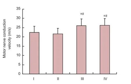
Figure 3 Effects of icariin on motor nerve conduction velocity in rats with sciatic nerve injury.

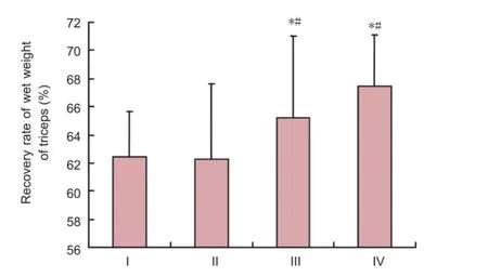
Figure 4 Icariin effects on motor percentage recovery (wet weight) of triceps muscle in rats with sciatic nerve injury.
Osmium tetroxide staining
Following electrophysiological testing, the conduit and sciatic nerve 5 mm distal and proximal to the conduit were dissected out. The sciatic nerve was fxed in 4% paraformaldehyde for 12 hours, rinsed briefy under running tap water, stained with 1% osmium tetroxide (Acros, Waltham, MA, USA) for 12 hours, and rinsed briefy again under running water. The tissue was then dehydrated through a graded alcohol series, permeabilized, embedded in paraffn, and sliced into transverse sections (5 µm thick). The sections were mounted, dried by baking, dewaxed, mounted in neutral resin, and viewed under a light microscope (Leica). Cross-sectional images of the nerve were obtained and the number of myelinated nerve fbers per visual feld (400 × magnifcation)was calculated using Image tool 3.0 software (Department of Dental Diagnostic Science at The University of Texas Health Science Center, San Antonio, TX, USA). Each section was counted three times, and the mean was calculated.

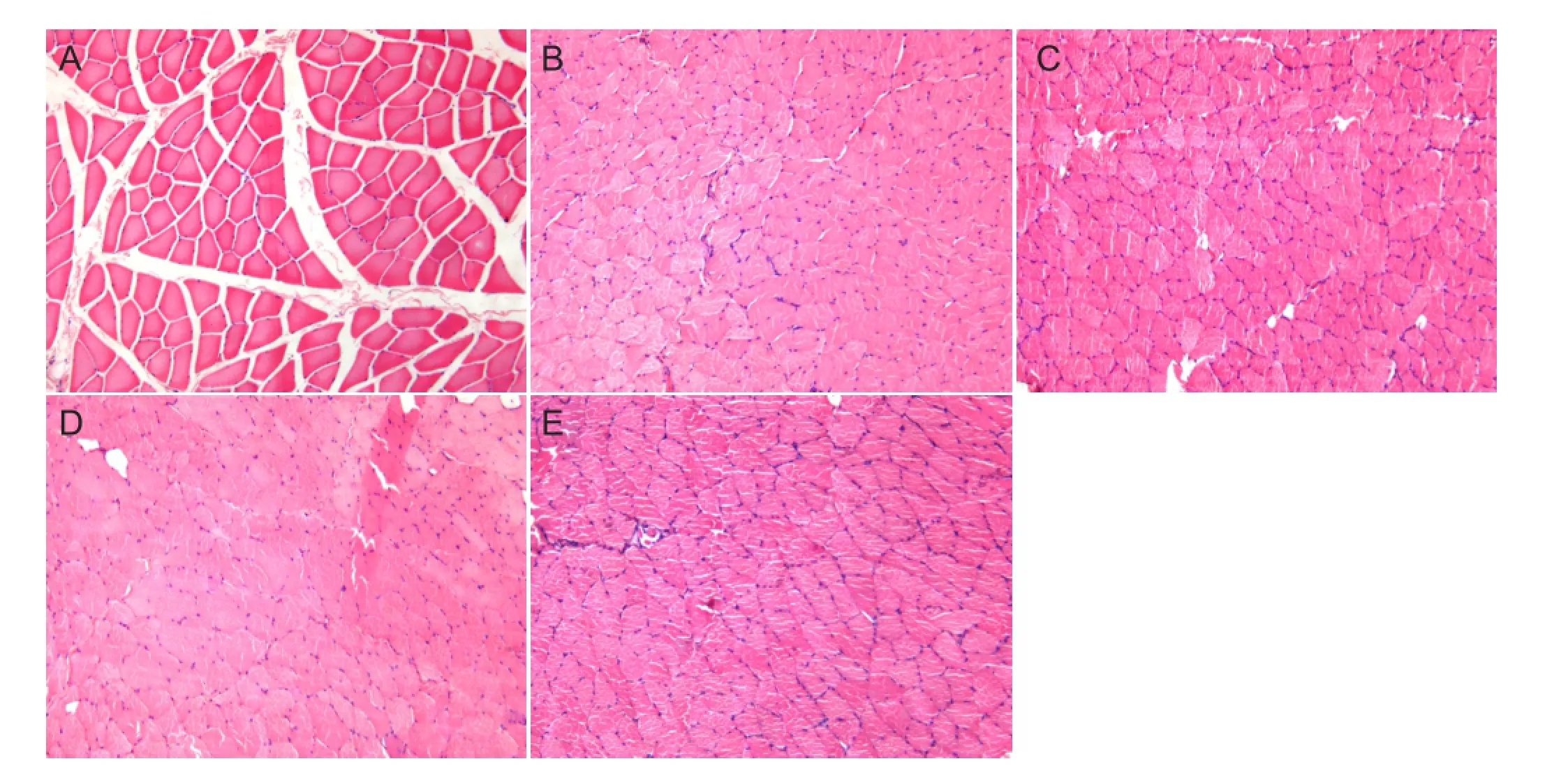
Figure 5 Effects of icariin and NGF on pathological changes in triceps of rats with sciatic nerve injury (hematoxylin-eosin staining, × 400).

Figure 6 Effects of icariin and NGF treatment on myelinated nerve fbers of rats with sciatic nerve injury.
Statistical analysis
Data were processed and analyzed using SPSS 13.0 software (SPSS, Chicago, IL, USA). All data were expressed as the mean ± SD. Intergroup comparisons were performed by oneway analysis of variance, and independent samples t-test. P <0.05 was considered statistically signifcant.
Results
General condition of rats with sciatic nerve injury
The rats were in good condition following surgery, with no observed wound infection (and therefore no rats excluded). In the physiological saline group, three rats experienced ulceration of the right hindpaw after surgery. Autophagy of the toes was observed at 12 weeks in the same three rats, and their ulcers had not healed. In the nerve growth factor solution group, four rats had ulceration of the right hindpaw. At 12 weeks, autophagy was noted in two of those rats, whereas ulcer healing was visible in the remaining two. In the icariin group, ulceration of the right hindpaw occurred in three rats. At 12 weeks, toe autophagy appeared in two of them, and ulcers had healed in the third rat. In the nerve growth factor sustained-release microsphere group, fve rats suffered from ulceration of the right hindpaw. At 12 weeks, toe autophagy was observed in three of those rats, and ulcer healing was detectable in the remaining two.
Sciatic functional index
In all groups, sciatic functional index showed a trend of gradual recovery with time. After 8 weeks, recovery slowed. No significant differences in sciatic functional index were detected between groups at any time point examined (P > 0.05;Figure 2).
Sciatic nerve conduction velocity
Electrophysiological tests revealed that at 12 weeks after surgery, sciatic nerve conduction velocity was greater in rats that had received icariin or nerve growth factor sustained-release microspheres than in those that received physiological saline or nerve growth factor solution (P < 0.05;Figure 3). Moreover, sciatic nerve conduction velocity in the icariin group was similar to that in the nerve growth factor sustained-release microsphere group (P > 0.05;Figure 3).
Triceps muscle weight
Twelve weeks after surgery, triceps muscles on the experimental side of rats in all groups were atrophic compared with the control side. The percentage recovery was signifcantly higher in the icariin and nerve growth factor sustained-release microsphere groups than in the physiological saline and nerve growth factor solution groups (P < 0.05), with no signifcant difference between the icariin and nerve growth factor sustained-release microsphere groups (P > 0.05;Figure 4).
Pathological changes in triceps muscle
After hematoxylin-eosin staining at 12 weeks after surgery, left (control) side triceps muscle fibers had clearly-defined boundaries, uniform staining, and regular diameter. On the right (experimental) side, muscle fbers were smaller in diameter than in the left side, but no notable differences in morphology were observed between the groups (Figure 5).
Pathological changes in the sciatic nerve
Osmium tetroxide staining 12 weeks after surgery revealed round or elliptical sciatic nerve fibers, and uniform nerve diameter and myelin sheath thickness in the left (control) side of rats from all four groups. However, in the right (experimental) side, nerve diameter and myelin sheath thickness were varied, and smaller than the control side. There were more myelinated nerve fbers in the icariin and nerve growth factor sustained-release microsphere groups than in the physiological saline group (P < 0.05). No signifcant differences in the number of myelinated nerve fibers were detected between the icariin and nerve growth factor sustained-release microsphere groups (P > 0.05;Figure 6).
Discussion
During peripheral nerve repair, neurons and Schwann cells secrete various factors that promote nerve regeneration (Ide, 1996). Nerve growth factor was the first such factor to be discovered and is the most extensively studied. It stimulates synapse growth, promotes differentiation, development and maturation of sympathetic and sensory neurons, and maintains normal neuronal functions (Aloe et al., 2012; Manni et al., 2013; Bothwell, 2014). Nerve growth factor solution and nerve growth factor sustained-release microspheres were therefore used as controls to icariin in the present study. Nerve growth factor is readily absorbed by tissues after direct transient topical application of nerve growth factor solution. However, peripheral nerve regeneration is a long process, with sciatic nerve repair taking at least 6 weeks in rats. Thus, a drug delivery system is advantageous in the promotion of peripheral nerve regeneration (Wang et al., 2014). Wood et al. (2011, 2013) showed that local application of poly (lactic-co-glycolic acid) sustained-release microspheres containing glial cell-derived neurotrophic factor could effectively improve the recovery of motor function in peripheral nerves during delayed repair. Takagi et al. (2012) demonstrated that composite biological sleeves obtained a better outcome when combined with local sustained-release brain-derived neurotrophic factor in bridging of a peripheral nerve defect. In our previous study, we successfully prepared nerve growth factor-poly (lactic-co-glycolic acid) sustained-release microspheres by water-oil-water multiple emulsion solvent evaporation. Our animal studies demonstrated that poly (lactic-co-glycolic acid) sustained-release microspheres containing nerve growth factor effectively elevated the number and maturity of axons and increased the effects of nerve repair in models after small gap bridging suture (Wang et al., 2014). Therefore, nerve growth factor sustained-release microspheres were used as positive controls in the present study.
Here, we found that icariin suspension exhibited identical promoting effects on nerve regeneration to nerve growth factor sustained-release microspheres, indicating that the absorption of icariin suspension in the conduit may also have a sustained-release function.
In summary, local application of icariin suspension contributes to peripheral nerve regeneration, increases the number of regenerating nerve fbers and nerve conduction velocity after sleeve bridging, and ultimately improves functional recovery. The outcomes of icariin suspension are similar to those of nerve growth factor sustained-release microspheres.
Author contributions:BC established animal models and detected sciatic nerve function. SPN performed animal histology staining and analyzed data. ZYW prepared drugs. ZWW made release microspheres. JXD provided literature data. PXZ, XFY and NH were in charge of manuscript authorization. YHK designed this study and wrote the manuscript. BGJ designed this study, analyzed data and was in charge of manuscript authorization. All authors approved the final version of the paper.
Conficts of interest:None declared.
Aloe L, Rocco ML, Bianchi P, Manni L (2012) Nerve growth factor: from the early discoveries to the potential clinical use. J Transl Med 10:239.
Bothwell M (2014) NGF, BDNF, NT3, and NT4. Handb Exp Pharmacol 220:3-15.
Chen CT, Lin JG, Lu TW, Tsai FJ, Huang CY, Yao CH, Chen YS (2010) Earthworm extracts facilitate PC12 cell differentiation and promote axonal sprouting in peripheral nerve injury. Am J Chin Med 38:547-560.
de Medinaceli L, Freed WJ, Wyatt RJ (1982) An index of the functional condition of rat sciatic nerve based on measurements made from walking tracks. Exp Neurol 77:634-643.
Gold BG (1997) Axonal regeneration of sensory nerves is delayed by continuous intrathecal infusion of nerve growth factor. Neuroscience 76:1153-1158.
Hare GM, Evans PJ, Mackinnon SE, Best TJ, Bain JR, Szalai JP, Hunter DA (1992) Walking track analysis: a long-term assessment of peripheral nerve recovery. Plast Reconstr Surg 89:251-258.
Höke A (2006) Mechanisms of disease: what factors limit the success of peripheral nerve regeneration in humans? Nat Clin Pract Neurol 2:448-454.
Hontanilla B, Aubá C, Gorría O (2007) Nerve regeneration through nerve autografts after local administration of brain-derived neurotrophic factor with osmotic pumps. Neurosurgery 61:1268-1275.
Hsiang SW, Lee HC, Tsai FJ, Tsai CC, Yao CH, Chen YS (2011) Puerarin accelerates peripheral nerve regeneration. Am J Chin Med 39:1207-1217.
Ide C (1996) Peripheral nerve regeneration. Neurosci Res 25:101-121.
Kou Y, Wang Z, Wu Z, Zhang P, Zhang Y, Yin X, Wong X, Qiu G, Jiang B (2013) Epimedium extract promotes peripheral nerve regeneration in rats. Evid Based Complement Alternat Med 2013:954798.
Lee AC, Yu VM, Lowe Iii JB, Brenner MJ, Hunter DA, Mackinnon SE, Sakiyama-Elbert SE (2003) Controlled release of nerve growth factor enhances sciatic nerve regeneration. Exp Neurol 184:295-303.
Ma H, He X, Yang Y, Li M, Hao D, Jia Z (2011) The genus Epimedium: an ethnopharmacological and phytochemical review. J Ethnopharmacol 134:519-541.
Manni L, Rocco ML, Bianchi P, Soligo M, Guaragna M, Barbaro SP, Aloe L (2013) Nerve growth factor: basic studies and possible therapeutic applications. Growth Factors 31:115-122.
Newman JP, Verity A, Hawatmeh S, Fee WE, Terris DJ (1996) CIliary neurotrophic factor enhances peripheral nerve regeneration. Arch Otolaryngol Head Neck Surg 122:399-403.
Quigley AF, Bulluss KJ, Kyratzis IL, Gilmore K, Mysore T, Schirmer KS, Kennedy EL, O’Shea M, Truong YB, Edwards SL, Peeters G, Herwig P, Razal JM, Campbell TE, Lowes KN, Higgins MJ, Moulton SE, Murphy MA, Cook MJ, Clark GM, et al. (2013) Engineering a multimodal nerve conduit for repair of injured peripheral nerve. J Neural Eng 10:016008.
Shindel AW, Xin ZC, Lin G, Fandel TM, Huang YC, Banie L, Breyer BN, Garcia MM, Lin CS, Lue TF (2010) Erectogenic and neurotrophic effects of icariin, a purifed extract of horny goat weed (Epimedium spp.) in vitro and in vivo. J Sex Med 7:1518-1528.
Takagi T, Kimura Y, Shibata S, Saito H, Ishii K, Okano HJ, Toyama Y, Okano H, Tabata Y, Nakamura M (2012) Sustained bFGF-release tubes for peripheral nerve regeneration: comparison with autograft. Plast Reconstr Surg 130:866-876
Tohda C, Nagata A (2012) Epimedium koreanum extract and its constituent icariin improve motor dysfunction in spinal cord injury. Evid Based Complement Alternat Med 2012:731208.
Utley DS, Lewin SL, Cheng ET, Verity A, Sierra D, Terris DJ (1996) Brain-derived neurotrophic factor and collagen tubulization enhance functional recovery after peripheral nerve transection and repair. Arch Otolaryngol Head Neck Surg 122:407-413.
Wang W, Huang CY, Tsai FJ, Tsai CC, Yao CH, Chen YS (2011) Growth-promoting effects of quercetin on peripheral nerves in rats. Int J Artif Organs 34:1095-1105.
Wang Z, Han N, Wang J, Zheng H, Peng J, Kou Y, Xu C, An S, Yin X, Zhang P, Jiang B (2014) Improved peripheral nerve regeneration with sustained release nerve growth factor microspheres in small gap tubulization. Am J Transl Res 6:413-421.
Wei S, Yin X, Kou Y, Jiang B (2009a) Lumbricus extract promotes the regeneration of injured peripheral nerve in rats. J Ethnopharmacol 123:51-54.
Wei S, Zhang P, Dang Y, Zhang H, Jiang B (2008) Primary study on effect of various components of modifed formula radix hedysari on peripheral nerve regeneration. Zhongguo Xiu Fu Chong Jian Wai Ke Za Zhi 22:1056-1059.
Wei SY, Zhang PX, Han N, Dang Y, Zhang HB, Zhang DY, Fu ZG, Jiang BG (2009b) Effects of Hedysari polysaccharides on regeneration and function recovery following peripheral nerve injury in rats. Am J Chin Med 37:57-67.
Wood MD, Kemp SW, Weber C, Borschel GH, Gordon T (2011) Outcome measures of peripheral nerve regeneration. Ann Anat 193:321-333.
Wood MD, Kim H, Bilbily A, Kemp SW, Lafontaine C, Gordon T, Shoichet MS, Borschel GH (2012a) GDNF released from microspheres enhances nerve regeneration after delayed repair. Muscle Nerve 46:122-124.
Wood MD, Gordon T, Kim H, Szynkaruk M, Phua P, Lafontaine C, Kemp SW, Shoichet MS, Borschel GH (2012b) Fibrin gels containing GDNF microspheres increase axonal regeneration after delayed peripheral nerve repair. Regen Med 8:27-37.
Wood MD, Gordon T, Kemp SW, Liu EH, Kim H, Shoichet MS, Borschel GH (2013) Functional motor recovery is improved due to local placement of GDNF microspheres after delayed nerve repair. Biotechnol Bioeng 110:1272-1281.
Xu H, Jiang B, Zhang D, Fu Z, Zhang H (2002) Compound injection of radix Hedysari to promote peripheral nerve regeneration in rats. Chin J Traumatol 5:107-111.
Xu X, Yee WC, Hwang PY, Yu H, Wan AC, Gao S, Boon KL, Mao HQ, Leong KW, Wang S (2003) Peripheral nerve regeneration with sustained release of poly(phosphoester) microencapsulated nerve growth factor within nerve guide conduits. Biomaterials 24:2405-2412.
Xue L, Wang Y, Jiang Y, Han T, Nie Y, Zhao L, Zhang Q, Qin L (2012) Comparative effects of er-xian decoction, epimedium herbs, and icariin with estrogen on bone and reproductive tissue in ovariectomized rats. Evid Based Complement Alternat Med 2012:241416.
Zhang P, Han N, Wang T, Xue F, Kou Y, Wang Y, Yin X, Lu L, Tian G, Gong X, Chen S, Dang Y, Peng J, Jiang B (2013) Biodegradable conduit small gap tubulization for peripheral nerve mutilation: a substitute for traditional epineurial neurorrhaphy. Int J Med Sci 10:171-175.
Copyedited by Slone-Murphy J, Stow A, Yu J, Qiu Y, Li CH, Song LP, Zhao M
*Correspondence to: Yu-hui Kou, M.D., yuhuikou@bjmu.edu.cn. Bao-guo Jiang, M.D., jiangbaoguo@vip.sina.com.
# These authors contributed equally to this work.
10.4103/1673-5374.150711
http://www.nrronline.org/
Accepted: 2014-11-04
- 中国神经再生研究(英文版)的其它文章
- Letter from the Editors-in-Chief
- Fat cell-secreted adiponectin mediates physical exercise-induced hippocampal neurogenesis: an alternative anti-depressive treatment?
- Induced pluripotent stem cell-derived neural stem cell therapies for spinal cord injury
- “Bad regenerators” die after spinal cord injury: insights from lampreys
- Can cinnamon bring aroma in Parkinson’s disease treatment?
- Acupuncture: a potent therapeutic tool for inducing adult neurogenesis

