Raptor对胶质瘤细胞侵袭能力的影响
张洪旺, 张宝刚, 宋瑞卉, 王汉秋, 郭文君
(潍坊医学院病理教研室,山东 潍坊 261053)
Raptor对胶质瘤细胞侵袭能力的影响
张洪旺, 张宝刚, 宋瑞卉, 王汉秋, 郭文君△
(潍坊医学院病理教研室,山东 潍坊 261053)
目的: 研究Raptor对于胶质瘤细胞侵袭能力的影响。方法: 采用RNA干扰技术,向胶质瘤U87细胞转染Raptor限定性siRNA干扰质粒, Western blotting检测转染后细胞Raptor的表达水平以鉴定转染效果。体外侵袭实验检测Raptor表达降低后,U87细胞侵袭能力的变化;Western blotting检测细胞中ARK5的磷酸化情况和MMP-2、MMP-9的表达水平。免疫组织化学法检测低级别及高级别胶质瘤中Raptor的表达水平。结果: 转染Raptor siRNA质粒的U87细胞命名为siRaptor/U87,转染对照组质粒的细胞命名为Scr/U87,转染成功的实验组细胞Raptor的表达降低。体外侵袭实验中siRaptor/U87较对照组穿透基质膜的细胞数少(P<0.01)。Western blotting显示实验组细胞中磷酸化ARK5、MMP-2和MMP-9蛋白的表达水平均较对照组低。胶质瘤组织中Raptor的表达与恶化程度存在相关性(P<0.01)。结论: Raptor 表达降低可能通过磷酸化ARK5及增加MMP-2、MMP-9的表达促进胶质瘤细胞的侵袭力。
Raptor蛋白; 胶质瘤; 肿瘤侵袭; 哺乳动物霉帕雷素靶蛋白; AMPK相关激酶5; 基质金属蛋白酶-2; 基质金属蛋白酶-9
Raptor是哺乳动物雷帕霉素靶蛋白(mammalian target of rapamycin,mTOR)的一种调控蛋白。1993年,Hara等[1]证实了mTOR于酵母菌属真菌细胞中的存在,1994年,Brown等[2]通过实验鉴定了mTOR(亦称FRAP、RAFT1或RAPT)基因及蛋白产物。mTOR属磷脂酰肌醇3-激酶(phosphatidylinositol 3-kinase,PI3K)蛋白激酶家族,是PI3K/PKB信号通路的下游效应蛋白,在细胞中有广泛而重要的作用。Raptor与mLst8、FKBP38、Deptor、PRAS40结合mTOR在细胞中以复合物形式存在,能够连接mTOR与下游效应蛋白,磷酸化S6激酶1(S6 kinase 1,S6K1)和真核生物起始因子4E结合蛋白1(eukaryotic initiation factor 4E binding protein 1,4E2BP1),调节细胞内蛋白质的表达,从而改变细胞的生长和增殖状态,推测其亦能影响胶质瘤细胞的侵袭能力。有研究表明,AMPK相关激酶 5(AMPK-related kinase 5,ARK5)可以通过基质金属蛋白酶2(matrix metalloproteinase-2,MMP-2)和MMP-9在胶质瘤的侵袭和转移中发挥重要作用,联系之前实验对ARK5蛋白的研究[3],可以帮助探究mTOR-Raptor在胶质瘤细胞侵袭中的机制。本实验通过RNA干扰技术降低胶质瘤细胞系Raptor的表达并观察细胞侵袭能力变化,检测ARK5及其下游蛋白表达水平,探求mTOR-Raptor复合物在胶质瘤侵袭和转移中的作用机制。
材 料 和 方 法
1 临床资料
收集潍坊医学院病理学教研室2008~2012年的胶质瘤蜡块标本共52例,均经病理证实,其中低级别胶质瘤(WTOⅠ、Ⅱ级)22例,高级别胶质瘤(WTO Ⅲ、Ⅳ级)30例。所有患者的临床及病理资料齐全,并且术前均未经放疗、化疗。
2 主要试剂
培养基及相关试剂购自HyClone;Raptor限定性siRNA质粒和对照组siRNA质粒购自Genescript;细胞转染试剂盒购自Invitrogen;24孔趋化小室购自康宁世纪公司;Matrivgel购自北京威格拉斯公司;人EGF购自R&D;抗Raptor、MMP-2和MMP-9抗体购自北京中衫金桥公司;抗p-ARK5和ARK5抗体购自Santa Cruz。
3 方法
3.1 细胞培养和处理 人类胶质瘤细胞系U87购自ATCC,细胞培养基为含10%胎牛血清的RPMI-1640,置37 ℃、5% CO2培养箱中进行无菌培养。在加入完全培养基转染之前24 h,将2×10⁵U87细胞种植于35 mm培养皿内。依据说明书使用Lipofectamine 2000试剂进行转染,siRNA Raptor目标片段为5’-TATTTGGTCGTCCAATCTCGT-3’[4],对照组转染插入SCR序列的siRNA质粒(SCR/U87)。转染的实验组和对照组细胞分别命名为siRaptor/U87和U87。培养72 h后,用Western blotting评价存活细胞Raptor的表达。
3.2 体外侵袭实验 按照文献所述方法进行Boyden小室侵袭实验[5]。小室碳酸脂膜(8 μm)表面铺上一层以1.5 g/L浓度配制的基底膜基质。将siRaptor/U87和对照组SCR/U87细胞分别悬浮于无血清培养基中,浓度为4×108/L,37 °C孵育30 min后加入上室。下室加入约300 μL趋化介质(RPMI-1640,0.1%牛血清白蛋白和25 mmol/L HEPES)。37 °C、5% CO2孵育24 h,对侵袭至膜下表面的细胞进行固定和染色。400倍光镜下计数5块预定区域内侵袭至基质膜滤膜底面的细胞数,取其平均值。所有实验重复3遍。
3.3 Western blotting实验 U87、SCR/U87和si-Raptor/U87细胞在无血清培养基中分别用10 μg/L的表皮生长因子刺激30 min,使用1×十二烷基硫酸钠样品缓冲液溶解,提取总蛋白,蛋白变性后经过SDS-PAGE、转膜、抗体孵育、显影得到蛋白印迹,分析ARK5的磷酸化情况。使用之前实验抽提的SCR/U87和siRaptor/U87细胞蛋白,再次进行 Western blotting实验,分析2组细胞中MMP-2和MMP-9表达水平。所有实验重复3次。
3.4 免疫组化实验 根据说明书利用链霉亲和素过氧化物酶免疫组化分析测定所有胶质组织标本Raptor表达。石蜡切片的染色程度同时基于染色的强度和阳性细胞的比例,由2个观察者独立评估计分[6]。单个细胞的染色强度以如下标准衡量:0(不染色);1(弱染色,淡黄色);2(中等染色,黄棕色);3(强染色、棕色)。阳性细胞比例以如下标准分级:0(没有阳性细胞);1(阳性细胞数< 10%);2(阳性细胞数10%~50%);3(阳性细胞数> 50%)。染色指数计算方式如下:染色指数=染色强度×染色阳性细胞比例。通过染色指数评估Raptor的表达(得分为0、1、2、3、4、6、7),以染色指数得分≥ 6标志肿瘤高表达Raptor,染色指数≤4标志肿瘤低表达Raptor。
4 统计学处理
统计分析使用SPSS 16.0软件。定量数据结果用均数±标准差(mean±SD)表示,计数资料采用χ2检验;计量资料采用两个独立样本的t检验;相关性采用Spearman相关分析。以P<0.05表示差异有统计学意义。
结 果
1 转染siRaptor 降低U87细胞中Raptor的表达
应用RaptorsiRNA质粒转染72 h,转染成功后的U87细胞Raptor蛋白表达降低,见图1。

Figure 1.The expression of Raptor in U87, siRaptor/U87 and SCR/U87 cells detected by Western blotting. β-actin was used as an internal reference.
图1 Western blotting法检测各组细胞中Raptor 的表达情况
2 降低U87细胞中Raptor的表达导致U87细胞侵袭能力下降
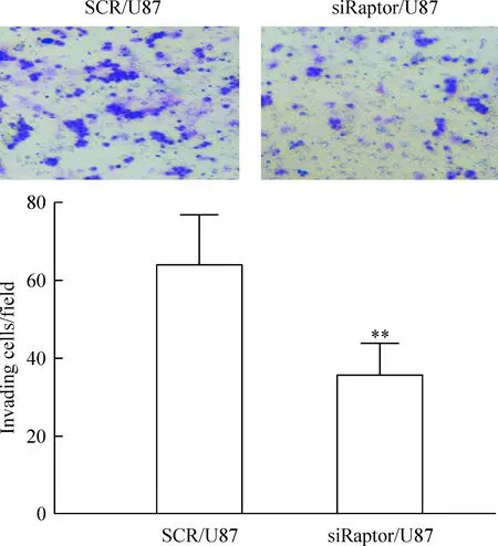
使用转染成功的siRaptor/U87细胞与对照组细胞进行侵袭实验,发现siRaptor/U87组比Scr/U87组穿透滤膜的细胞数减少,差异显著(P<0.01),见图2。

Figure 2.The effect ofRaptorknockdown on invasion ability of U87 cells (×200). Mean±SD.n=3.**P<0.01vsSCR/U87.
图2 干扰Raptor的表达对U87细胞侵袭能力的影响
3 siRaptor/U87细胞中ARK5的磷酸化水平
结果显示,siRaptor/U87较对照组细胞ARK5的磷酸化减少,见图3。

Figure 3.The expression of p-ARK5 protein and ARK5 protein in siRaptor/U87 and SCR/U87 detected by Western blotting.
图3 Western blotting法检测siRaptor/U87与对照组细胞中ARK5的磷酸化水平
4 siRaptor/U87细胞中MMP-2和MMP-9的表达
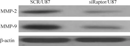
结果显示,siRaptor/U87较对照组细胞MMP-2和MMP-9的表达下调,见图4。

Figure 4.The expression of MMP-2 and MMP-9 in siRaptor/U87 cells and SCR/U87 detected by Western blotting. β-actin as an internal reference.
图4 Western blotting法检测siRaptor/U87与对照组细胞中MMP-2和MMP-9的表达情况
5 胶质瘤组织中Raptor蛋白的表达
免疫组织化学染色的52例胶质组织在显微镜下观察,正常胶质组织没有或极少表达Raptor,恶化程度高的胶质瘤细胞Raptor的表达要高于恶化程度低的胶质瘤细胞,见图5。统计分析显示,Raptor的表达与胶质瘤是否发生恶性细胞变化具有相关性(P<0.01),见表1。
讨 论
胶质瘤是中枢神经系统常见的一类肿瘤,其平均生存时间在12~15个月之间[7],被称为人类最具危害性的肿瘤。而肿瘤恶性程度与肿瘤细胞的侵袭有关,因此研究胶质瘤细胞的侵袭对提高治疗功效具有重要的作用[8-9]。Raptor具有潜在的原癌基因特性,在人类乳腺癌、前列腺癌[10]中高表达但尚未见Raptor与胶质瘤侵袭相关研究。明确Raptor在胶质瘤侵袭中的作用,对抑制胶质瘤的侵袭具有重要作用。同时实验证实,Raptor在胶质瘤中的表达具有正相关性,即:Raptor高表达时,通过RNA 干扰技术将相关质粒转染胶质瘤,培养72 h后,转染成功后的U87细胞Raptor蛋白表达降低。为研究Raptor与ARK5之间的关系我们进行了分子生物学实验,通过RNA 干扰技术使用小干扰RNA质粒转染乳腺癌细胞株,Western blotting 检测到Raptor表达降低,成功构建了稳定沉默Raptor表达的细胞系。体外癌细胞侵袭实验证明Raptor参与了癌细胞的侵袭过程。Raptor降低表达的实验组细胞ARK5的磷酸化减少,MMP-2和MMP-9的表达相应降低。通过免疫组化实验表明:Raptor在高级胶质瘤(WTO分级为Ⅲ、Ⅳ)中的表达明显高于在低级胶质瘤(WTO分级为Ⅰ、Ⅱ)中的表达。以上实验结果表明,Raptor可以磷酸化ARK5,上调MMP-2和MMP-9的表达,从而调控胶质瘤细胞的侵袭。对侵袭机制的研究可为胶质瘤的防治提供参考,为明确Raptor在胶质瘤发展中的作用及与侵袭相关的下游信号分子之间的相互关系,我们将进行进一步实验。

Figure 5.The expression of Raptor in tissues of grade I, grade II, grade III and grade IV gliomas(×400).
图5 I、II、III和IV级胶质瘤组织中Raptor的表达
表1 Raptor的表达与胶质瘤恶化程度的关系
Table 1.The relationship between Raptor expression and degree of deterioration of glioma

WTOclassificationgradenRaptorHighPositiverateNegativeorexpression(%)lowexpressionI,II221560.826III,IV3022**82.098
**P<0.01vsI,II.
[1] Hara K, Maruki Y, Long X, et al. Raptor, a binding partner of target of rapamycin (TOR), mediates TOR action[J]. Cell, 2002, 110(2):177-189.
[2] Brown EJ, Albers MW, Shin TB, et al. A mammalian protein targeted by G1-arresting rapamycin-receptor complex[J]. Nature,1994, 369(6483):756-758.
[3] Kusakai G, Suzuki A, Ogura T, et al. ARK5 expression in colorectal cancer and its implications for tumor progression [J]. Am J Pathol, 2004, 164(3):987-995.
[4] Chen P, Li K, Liang Y, et al. High NUAK1 expression correlates with poor prognosis and involved in NSCLC cells migration and invasion[J]. Exp Lung Res, 2013, 39(1):9-17.
[5] Baik SH, Kim NK, Lee KY, et al. Factors influencing pathologic results after total mesorectal excision for rectal cancer: analysis of consecutive 100 cases[J]. Ann Surg Oncol, 2008, 15(3):721-728.
[6] Li J, Guan HY, Gong LY, et al. Clinical significance of sphingosine kinase-1 expression in human astrocytomas progression and overall patient survival[J]. Clin Cancer Res, 2008, 14(21):6996-7003.
[7] Stupp R, Hegi ME, Gilbert MR, et al. Chemoradiotherapy in malignant glioma: standard of care and future directions[J]. J Clin Oncol, 2007, 25(26):4127-4136.
[8] Huveldt D, Lewis-Tuffin LJ, Carlson BL, et al. Targeting Src family kinases inhibits bevacizumab-induced glioma cell invasion[J].PLoS One, 2013, 8(2):e56505.
[9] 王 影,孙 静,李艳艳. miR-29a对胶质瘤细胞CDC42 表达及迁移和侵袭的影响[J]. 中国肿瘤临床,2012, 40 (11):629-633.
[10]Lu S, Niu N, Guo H, et al. ARK5 promotes glioma cell invasion, and its elevated expression is correlated with poor clinical outcome[J]. Eur J Cancer, 2013, 49(3):752-763.
Effect of Raptor on invasion ability of glioma cells
ZHANG Hong-wang, ZHANG Bao-gang, SONG Rui-hui, WANG Han-qiu, GUO Wen-jun
(DepartmentofPathology,WeifangMedicalUniversity,Weifang261053,China.E-mail: 13963663745@163.com)
AIM: To study the influence of Raptor on the invasion ability of glioma cells. METHODS: The technique of RNA interference was used. U87 cells were transfected with Raptor restricted siRNA plasmid, and the expression level of Raptor in the transfected cells was detected by Western blotting. The invasive ability of the cancer cellsinvitrowas determined. The phosphorylation level of ARK5 and the expression of MMP-2 and MMP-9 were detected by Western blotting. The expression levels of Raptor in the tumor samples of low-grade gliomas (WTO grade I and grade II) and high-grade gliomas (WTO grade III and grade IV) were also analyzed by immunohistochemical staining. RESULTS:RaptorsiRNA was transfected into U87 cells and the cells were named siRaptor/U87 cells. The cells transfected with the control plasmid was named Scr/U87 cells. The expression level of Raptor in siRaptor/U87 cells was lower than that in Scr/U87 cells. The results of invitroinvasion assay showed that the number of siRaptor/U87 cells penetrating the Matrivgel matrix membrane was less than that of Scr/U87 cells (P<0.01). The protein expression of MMP-2 and MMP-9, and phosphorylation of ARK5 protein in the cells in the experimental group were lower than those in control group. The correlation between the expression of Raptor in gliomas and the degree of deterioration was also observed (P<0.01). CONCLUSION: The expression of Raptor may contribute to the invasion ability of glioma cells by phosphorylation of ARK5 and increase in the levels of MMP-2 and MMP-9.
Raptor protein; Glioma; Neoplasm invasiveness; Mammalian target of rapamycin; AMPK-related kinase 5; Matrix metalloproteinase-2; Matrix metalloproteinase-9
1000- 4718(2014)12- 2280- 04
2014- 06- 06
2014- 10- 16
R730.23
A
10.3969/j.issn.1000- 4718.2014.12.030
△通讯作者 Tel: 0536-8068957; E-mail: 13963663745@163.com

