Role of 3.0 T dynamic contrast-enhanced MRA of the breast in differentiating benign and malignant lesions
SHEKHAR Kumar Mehta,LIU Wan-hua
(Department of Radiology,Zhongda Hospital,Southeast University,Nanjing 210009,China)
Breast MRI(bMRI)is the most accurate method for detection of breast cancer with sensitivity that is nearly 100%,with lower specificity rates[1-7].Recently,MR angiography(MRA)of the breast has been used in characterizing breast lesions.Dynamic contrast-enhanced images of the breast are post-processed in different views by image subtraction and then typical maximum intensity projection(MIP)images are obtained.MIP images reveal not only the presence of enhancing lesions but also the angiographic vascular map of vessels within the breast.However,differentiation between arterial and venous breast vessels was not possible and hence was evaluated in general without differentiation of arteries and veins.
In our study,we first evaluated ipsilateral increased vascularity(difference between ipsilateral vessels and contralateral vessels)and secondly,we evaluated presence of adjacent vessel sign in breast lesions.The ipsilateral higher vascularity is believed to be due to neoangiogenic stimulation of the whole breast by breast cancer,reduced flow resistance in the tumor vessels,tumors higher metabolism or a combination of these factors[8-9].
1 Materials and Methods
1.1 Subjects
From December 2010 to February 2012,108 patients(52 malignant and 56 benign lesions)with unilateral and histopathologically confirmed breast lesions were included in this study(aged 27-80 years;mean 46 years).Patients with bilateral breast lesions,with a history of radiation therapy or breast biopsy within 6 months or unilateral mastectomy for a previous breast cancer or with no histologic confirmation of lesions were excluded.
Out of 52 patients with breast malignancies:38 were invasive ductal carcinoma(IDC),3 ductal carcinoma insitu(DCIS),8 DCIS with IDC,2 invasive lobular carcinoma(ILC)and 1 metastatic malignant melanoma.Out of 56 benign lesions:9 fibroadenoma with adenosis,8 only fibroadenoma,12 only adenosis,3 adenosis with apocrine metaplasia,7 fibrocystic breast disease,3 papilloma,1 calcified nodule,1 lipoma,1 ductal epithelial dysplasia,1 phyllode tumor and 10 lesions were associated with inflammatory lesions.
1.2 MRI protocol
MRI was performed with a 3-T MRI unit(Siemens).Patients were placed in prone position and both breasts imaged simultaneously using a double breast coil.MRI was done during 7-14th days of menstrual cycle.0.1 mmol·kg-1of gadopentetate dimeglumine(Gd-DTPA,Magnevist)was used intravenously at the rate of 2 ml·s-1followed by 10 ml of saline flush.Dynamic contrastenhanced image acquisition was started immediately 25 seconds after the bolus injection and series of 8 post contrast dynamic images were obtained at 1,2,3,4,5,6,7 and 8 min respectively.
For generation of MRA,unenhanced images in the dynamic sequence were subtracted from the second series of contrast-enhanced images,and MIPreconstruction was applied to the subtracted images.
1.3 Analysis of MR images
Coronal and transverse MIPs were prepared from the subtracted MR images and 3D rotation on workstation was used for assessment of vascularity of breast lesions.
According to the method defined by Sardanelli et al[8],the number of vessels,including both arteries and veins,3 cm or longer and 2 mm or greater in maximal diameter were counted for each breast.Absence of vessels at least 3 cm long and 2 mm in diameter indicated no or very low vascularity with score 0;presence of only one such vessel indicated low vascularity with score 1;presence of two to four such vessels indicated moderate vascularity with score 2;and the presence of five or more such vessels indicated high vascularity with score 3.Vascularity was considered increased when the difference in the number of vessels was two or greater in the breast containing the lesion compared with the contralateral breast.Vessels more than 3 mm in diameter within the breast tissue were considered enlarged,and the presence of such vessels was recorded.The presence of vessels either entering the enhancing lesion or in contact with the lesion edge on MIP images was accepted as presence of the adjacent vessel sign.
1.4 Statistical analysis
Microsoft-excel 2010 and SPSS18 for Windows were used for data collection and analysis.The number of vessels per breast in malignant and benign lesions was compared by the use of the independent two-sample Student's t test after the distribution of the number of vessels were tested with the Kolmogorov-Smirnov test.Two-sided Spearman's test was applied to test lesions and its association with ipsilateral increased vascularity and the adjacent vessel sign.A value of P<0.05 was considered to indicate a significant difference.The sensitivity,specificity,positive predictive value,negative predictive value,and accuracy of ipsilateral increased vascularity and the adjacent vessel sign were calculated.
2 Results
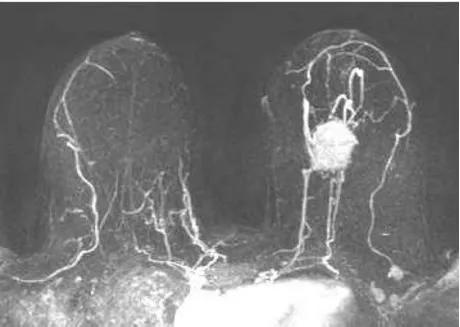
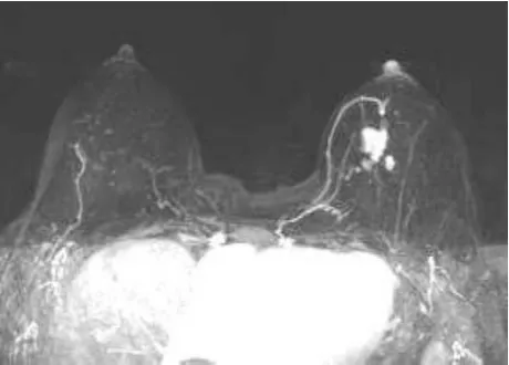
The adjacent vessel sign was found in 64(59%)patients(46 malignant and 18 benign lesions)out of the total 108 patients under study group.Of which,46 lesions(89%)out of 52 malignant lesions were true positive(TP,Fig 1)lesions and the remaining 6(11%)were false negative lesions.18(32%)out of 56 benign lesions were false positive(FP,Fig 2)and the remaining 38(68%)were true negative(TN)lesions.The sensitivity and specificity of adjacent vessel sign for indicator of malignancy were 89%and 68%respectively with positive predictive value(PPV)and negative predictive value(NPV)being 72%and 87%respectively(Tab 1).

Fig 1 A 52 year-old woman with invasive ductal carcinoma(mass lesion)of the left breast Maximum-intensity-projection reconstructed image of breast shows true-positive findings with both ipsilateral increased vascularity and adjacent vessels sign

Fig 2 A 48 year-old woman with fibroadenoma of the left breast Maximum-intensity-projection reconstructed image of breast shows false-positive findings of ipsilateral increased vascularity and adjacent vessels sign

Tab 1 Sensitivity,specificity,positive and negative predictive values and accuracies of ipsilateral increased vascularity and adjacent vessel sign as an indicator of ipsilateral cancer
Ipsilateral increased vascularity was found in 50(47%)patients(41 malignant and 9 benign)out of the total 108 patients who underwent contrast-enhanced MRI.Breast malignancy was present in 41 of 52 patients(79%)with ipsilateral increased vascularity.These cases were true-positive(TP,Fig 1).In 9 of the 50 patients with ipsilateral increased vascularity,the diagnosis was benign(18%)and was false positive(FP,Fig 2)cases.In 11 of the 52 breast cancer patients(21%),ipsilateral increased vascularity was not found and was false negative(FN)cases.In 47 of the 56 patients with benign lesions,no ipsilateral increased vascularity was found and was true negative(TN)cases.The sensitivity and specificity of ipsilateral increased vascularity as an indicator of malignancy were 79%and 84%with positive predictive value(PPV)and negative predictive value(NPV)being 71%and 86%respectively(Tab 1).
The mean and score of adjacent vessels,ipsilateral vessels,contralateral vessels of malignant and benign lesions were calculated(Tab 2).

Tab 2 Comparison of mean and score of adjacent vessels,ipsilateral vessels,contralateral vessels and ipsilateral increased vessels between malignant and benign lesions
3 Discussion
Breast MRA can be used as an integral part in addition to other breast MRI methods to increase diagnostic specificity.MIP images reveal not only the presence of enhancing lesions but also the angiographic vascular map of vessels within the breast by postprocessing the subtracted images in different views as malignancies are believed to undergo angiogenesis resulting in increased production of new blood vessels.
Using the method described by Sardanelli et al.[8,10],all the main vessels and their branches which were 3 cm or longer in length and 2 mm or wider in diameter for ipsilateral vessels,contralateral vessels were counted by freely rotating transverse and coronal MIP images at workstation.As shown previously by Kuhl et al.[9]that ipsilateral increased vascularity and adjacent vessel sign were highly associated with ipsilateral breast malignancies.Mahfouz and Sardanelli et al.[8,11]also showed an association between breast cancer and a higher ipsilateral vascularity.Our study generated similar results with ipsilateral increased vascularity and adjacent vessel sign seen more often in breast malignancies than benign lesions.
There was also significant difference in total number of adjacent vessels,ipsilateral vessels and contralateral vessels in breast bearing malignancies as compared to that of benign lesions(P<0.05).Possible explanation is higher angiogenetic activity in malignant cases,which ultimately results in increased global vascularity and presence of adjacent vessel sign.Small number of cases was main limitation of this study.However,it can be assumed that evaluation of ipsilateral increased vascularity and adjacent vessel sign can be a helpful integral in characterizing breast lesions in combination with other breast MRI methods.
4 Acknowledgements
I am very grateful and thankful to my Prof.Dr.LIU Wan-hua for reviewing the manuscript of article and providing valuable suggestions throughout the research.I would also like to thank my colleague Dr.WANG Rui for helping with data collection and statistical advice and Mrs.SUSHMA Singh for computer related works.
[1]FISCHER D R,MALICH A,WURDINGER S,et al.The adjacent vessel on dynamic contrast-enhanced breast MRI[J].AJR Am JRoentgenol,2006,187:W147-W151.
[2]FISCHER D R,WURDINGER S,BOETTCHER J,et al.Further signs in the evaluation of magnetic resonance mammography:a retrospective study[J].Invest Radiol,2005,40:430-435.
[3]MALICH A,FISCHER D R,WURDINGER S,et al.Potential MRI interpretation model:differentiation of benign from malignant breast masses[J].AJR Am JRoentgenol,2005,185:964-970.
[4]PETERSN H,BOREL-RINKESI H,ZUITHOFF N P,et al.Meta-analysis of MR imaging in the diagnosis of breast lesions[J].Radiology,2008,246:116-124.
[5]LEE S H,CHO N,KIM S J,et al.Correlation between high resolution dynamic MR features and prognostic factors in breast cancer[J].Korean JRadiol,2008,9:10-18.
[6]KO E Y,HAN B K,SHIN JH,et al.Breast MRI for evaluating patients with metastatic axillary lymph node and initially negative mammography and sonography[J].Korean J Radiol,2007,8:382-389.
[7]KIM DY,MOON WK,CHON,et al.MRI of the breast for the detection and assessment of the size of ductal carcinoma in situ[J].Korean J Radiol,2007,8:32-39.
[8]SARDANELLI F,IOZZELLI A,FAUSTO A,et al.Gadobenate dimeglumine-enhanced MR imaging breast vascular maps:association between invasive cancer and ipsilateral increased vascularity[J].Radiology,2005,235:791-797.
[9]SIBEL K,AYSEGÜL C,ETEM A,et al.Contrast-enhanced MR angiography of the breast:evaluation of ipsilateral increased vascularity and adjacent vessel sign in the characterization of breast lesions[J].AJR Am JRoentgenol,2010,195:1250-1254.
[10]SARDANELLI F,FAUSTO A,MENICAGLI L,et al.Breast vascular mapping obtained with contrast enhanced MR imaging:implications for cancer diagnosis treatment and risk stratification[J].Eur Radiol,2007,17(suppl 6):48-51.
[11]MAHFOUZ A E,SHERIF H,SAAD A,et al.Gadolinium-enhanced MR angiography of the breast:is breast cancer associated with ipsilateral higher vascularity?[J].Eur Radiol,2001,11:965-969.
